|
※サムネイル画像をクリックすると拡大画像が表示されます。
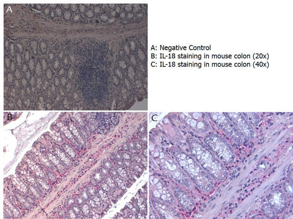
Immunohistochemistry with Rabbit anti-Mouse IL-18 antibody showing IL-18 staining in inflammatory cells of the mucous corium of mouse colon at 20x and 40x. Slide A is a negative control. Slides B and C show staining. Formalin fixed/paraffin embedded sections were subjected to heat induced epitope retrieval (HIER) at pH 6.2 and then incubated with mouse anti-IL-18 antibody at 4.0 μg/ml for 60 minutes. The reaction was developed using MACH 4 universal AP polymer detection system and visualized with WARP RED.
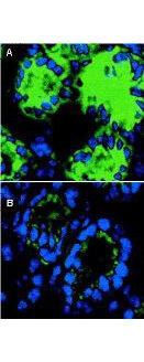
Immunofluorescence microscopy of IL-18 in mouse colon sections. The transversing portion of the large intestine from DSS-exposed (Panel A) and -unexposed mice (Panel B) was excised, rinsed in PBS, and frozen on isopentane cooled with liquid nitrogen. Frozen sections (5 μm) were cut on a Leica CM 1850 cryostat. The slides were fixed for 10 min in 4% paraformaldehyde, air-dried, and incubated for 20 min in PBS supplemented with 10% normal goat serum. Sections were incubated in a 1:50 dilution of Rockland's rabbit anti-Mouse IL-18 antibody or 1 μg/ml nonimmune rabbit IgG (not shown) as negative control. The antibodies were diluted in PBS containing 1% bovine serum albumin. After an overnight incubation at 4°C, the sections were washed three times with 0.5% bovine serum albumin in PBS. The sections were then incubated with a secondary goat anti-rabbit antibody conjugated to Alexa488 (Molecular Probes) for 60 min at room temperature in the dark. Nuclei were counterstained blue using 1 μg/100 ml bisbenzimide. After staining, sections were washed and examined with the Leica DM RXA confocal laser scanning system and analyzed. Similar staining will occur with other systems.
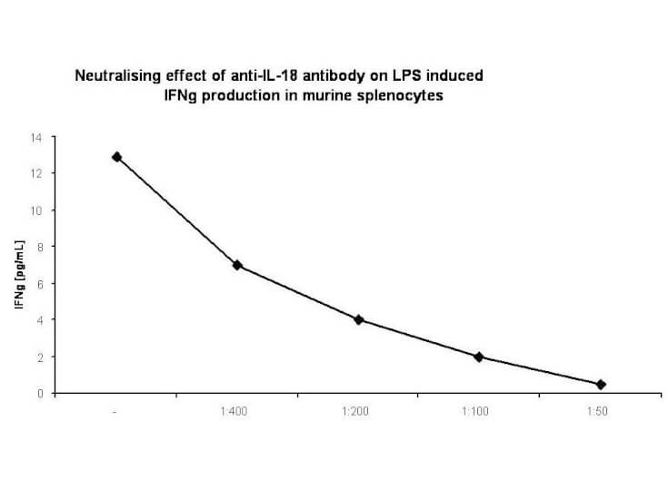
In-vitro neutralization. Spleens were aseptically removed and cell suspensions were prepared. Cells were washed twice and resuspended in RPMI supplemented with 10% FBS. For cytokine measurement, spleen cells were cultured at 5 mln/mL in 24-well, flat-bottom culture plates in the presence of several dilutions of rabbit anti-murine IL-18 antibody (1:400; 1:200; 1:100; 1:50) and 100 ng/mL of LPS (a phenol-extracted preparation from Escherichia coli 055:B5, Sigma Chemical Co). Cultures were incubated at 37°C in a humidified atmosphere with 5% CO2. At the end of the incubation period, cultures were frozen at -70°C and subjected to 3 freeze-thaw cycles to obtain total cytokine levels. Before assaying, samples were centrifuged for 10 minutes at 10,000g to remove debris.
|
|
|
|
Immunohistochemistry with Rabbit anti-Mouse IL-18 antibody showing IL-18 staining in inflammatory cells of the mucous corium of mouse colon at 20x and 40x. Slide A is a negative control. Slides B and C show staining. Formalin fixed/paraffin embedded sections were subjected to heat induced epitope retrieval (HIER) at pH 6.2 and then incubated with mouse anti-IL-18 antibody at 4.0 μg/ml for 60 minutes. The reaction was developed using MACH 4 universal AP polymer detection system and visualized with WARP RED.
|
|
| 別品名 |
rabbit anti-IL-18 antibody, rabbit anti-interleukin-18 antibody, Iboctadekin antibody, IFN gamma inducing factor antibody, IGIF antibody, IL 1 gamma antibody, IL 18 antibody, IL 1g antibody, IL-1F4, IL 18, Interleukin 18, IL18, Interleukin18, IL1 F4, IL1F4, Il18
|
| 交差種 |
Mouse
|
| 適用 |
Immunohistochemistry
Immuno Fluorescence
|
| 免疫動物 |
Rabbit
|
| 標識物 |
Unlabeled
|
| 精製度 |
Ig fraction - Protein A
|
| GENE ID |
16173
|
| Accession No.(Gene/Protein) |
P70380.2, P70380
|
| Gene Symbol |
Il18
|
| 参考文献 |
[Pub Med ID]27016579
|
| [注意事項] |
濃度はロットによって異なる可能性があります。メーカーDS及びCoAからご確認ください。
|
|
| メーカー |
品番 |
包装 |
|
RKL
|
210-401-323
|
500 UG
|
※表示価格について
| 当社在庫 |
なし
|
| 納期目安 |
約10日
|
| 保存温度 |
-20℃
|
|
※当社では商品情報の適切な管理に努めておりますが、表示される法規制情報は最新でない可能性があります。
また法規制情報の表示が無いものは、必ずしも法規制に非該当であることを示すものではありません。
商品のお届け前に最新の製品法規制情報をお求めの際はこちらへお問い合わせください。
|
※当社取り扱いの試薬・機器製品および受託サービス・創薬支援サービス(納品物、解析データ等)は、研究用としてのみ販売しております。
人や動物の医療用・臨床診断用・食品用としては、使用しないように、十分ご注意ください。
法規制欄に体外診断用医薬品と記載のものは除きます。
|
|
※リンク先での文献等のダウンロードに際しましては、掲載元の規約遵守をお願いします。
|
|
※CAS Registry Numbers have not been verified by CAS and may be inaccurate.
|
