|
※サムネイル画像をクリックすると拡大画像が表示されます。
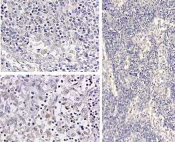
Immunohistochemistry of Human IL1 beta antibody. Tissue: human medullary lymph node. Fixation: formalin fixed paraffin embedded. Antigen retrieval: user optimized. Primary antibody: Human IL1 beta antibody. Secondary antibody: Peroxidase goat anti-rabbit at 1:10,000 for 45 min at RT. Localization: cytoplasm. Staining: Close up of medullary lymph node: positive staining in the cytoplasm of circulating macrophages. Neg Ctr (far right) of normal rabbit IgG with pH 6.2 at 40X.
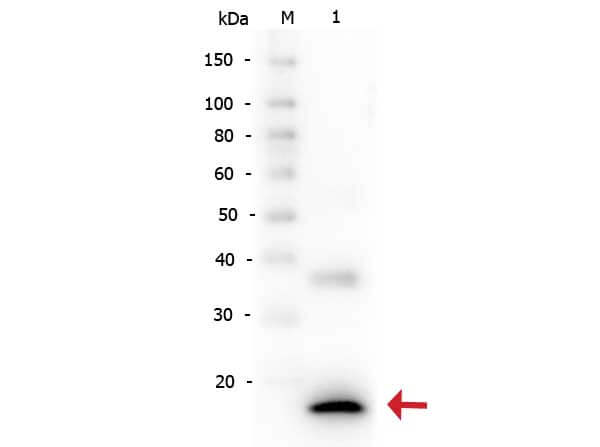
Western Blot of Rabbit anti-Human IL-1s antibody. Lane 1: Human IL-1s. Load: 5 ng per lane. Primary antibody: Human IL-1s antibody at 1:2,000 for overnight at 4°C. Secondary antibody: Peroxidase rabbit secondary antibody at 1:40,000 for 30 min at RT. Block: Blocking Buffer for Fluorescent Western Blotting (MB-070) for 30 min at RT. Predicted/Observed size: 17 kDa, 17 kDa for Human IL-1s. Other band(s): Unspecific band at ~35 kDa.
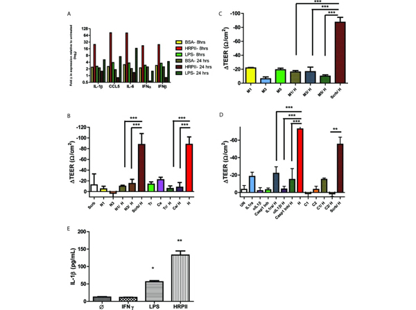
HRPII activates an inflammatory pathway in human cerebral microvascular endothelial cells. (A) qRT-PCR of chemokine/cytokine mRNA levels of hCMEC/D3 cells treated with 25?μg HRPII or BSA for 8?h and 24?h. (B) TEER measurements for?in vitro?hCMEC/D3 barriers transfected with shRNAs for NFκB (N1 and N3) or a scrambled control (Scrb) for 36?h or incubated with inhibitors for NFκB, celastrol (Ce), and triptolide (tr) for 2?h prior to addition of HRPII (H; 10?μg). Data are mean values ± SEM of results from 6 to 8 replicates pooled from three independent experiments. ***,?P?< 0.0001 (by one-way ANOVA). (C) TEER measurements for?in vitro?barriers transfected with shRNAs to MyD88 (M1 and M3 and M5) or a scrambled control (Scrb) for 36?h prior to addition of recombinant purified HRPII (10?μg). Data are mean ± SEM of results from 6 to 8 replicates pooled from 3 independent experiments. ***,?P?< 0.0001 (by one-way ANOVA). Results of assessment of knockdown levels are shown in?Fig.?S2?in the supplemental material. (D) TEER measurements for?in vitro?barriers transfected with shRNAs for caspase-1 (C1 and C2) or a scrambled control (Scrb) for 36?h or with IL-1Ra (500?ng), anti-IL-1β (αIL-1β) (25?ng), or the caspase-1 inhibitor YVAD-CMK (80?μM) (C1 Inh) for 1?h prior to treatment with recombinant purified HRPII (10?μg; H). Data are mean values ± SEM of results from 6 to 8 replicates pooled from four independent experiments. ***,?P?< 0.001 (by one-way ANOVA); **,?P?< 0.05 (by one-way ANOVA). (E) Quantitative ELISA for cleaved IL-1β from cell lysates. Cells were treated for 24?h with HRPII (10?μg), LPS (3?μg/ml), or IFN-γ (100?ng/ml) or left untreated. Data represent results from three biological replicates, each performed in triplicate. *,?P?= 0.0002; **,?P?= 0.0005 (compared to untreated control by unpaired?t?test). Fig 3. PMID:?27273825
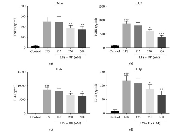
The effect of UK on the expressions of proinflammatory cytokines in LPS- stimulated BV-2?cells. Cells were pretreated with UK (125?nM, 250?nM, and 500?nM) for 12?h and with LPS (1000?ng/mL) treatment for another 12?h. The production of TNFα, PGE2, IL-6, and IL-1β?was measured by ELISA kits.?p?< 0.001 versus the control group,??p?< 0.05 versus the LPS-stimulated group,???p?< 0.01 versus the control group, and????p?< 0.001 versus the LPS-stimulated group,?n?=?3. Fig 2. PMID:?31428213
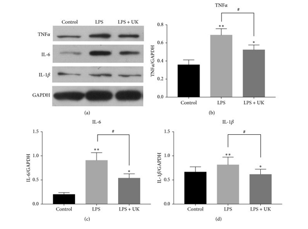
The effect of UK on the protein expressions of proinflammatory cytokines by western blot analysis. Cells were pretreated with UK (500?nM) for 12?h and with LPS (1000?ng/mL) treatment for another 12?h. The production of TNFα, IL-6, and IL-1β?was analyzed by western blot.?#p?< 0.05 versus the LPS-stimulated group,??p?< 0.05 versus the control group, and???p?< 0.01 versus the control group,?n?=?3. Fig 3. PMID:?31428213
|
|
|
|
Immunohistochemistry of Human IL1 beta antibody. Tissue: human medullary lymph node. Fixation: formalin fixed paraffin embedded. Antigen retrieval: user optimized. Primary antibody: Human IL1 beta antibody. Secondary antibody: Peroxidase goat anti-rabbit at 1:10,000 for 45 min at RT. Localization: cytoplasm. Staining: Close up of medullary lymph node: positive staining in the cytoplasm of circulating macrophages. Neg Ctr (far right) of normal rabbit IgG with pH 6.2 at 40X.
|
|
| 別品名 |
rabbit anti-IL-1 beta Antibody, rabbit anti-interleukin-1 beta antibody, IL-1 beta, Interleukin-1 beta, IL-1β, catabolin
|
| 交差種 |
Human
|
| 適用 |
Western Blot
Enzyme Linked Immunosorbent Assay
Immunohistochemistry
|
| 免疫動物 |
Rabbit
|
| 抗原部位 |
N-terminus
|
| 標識物 |
Unlabeled
|
| 精製度 |
Ig fraction - Ion Exchange /Gel Filtration
|
| GENE ID |
3553
|
| Accession No.(Gene/Protein) |
P01584.2, P01584
|
| Gene Symbol |
IL1B
|
| 参考文献 |
[[Cosmo Reference Collection]][Pub Med ID]27273825,31428213
|
| [注意事項] |
濃度はロットによって異なる可能性があります。メーカーDS及びCoAからご確認ください。
|
|
| メーカー |
品番 |
包装 |
|
RKL
|
209-401-301
|
1 MG
|
※表示価格について
| 当社在庫 |
なし
|
| 納期目安 |
約10日
|
| 保存温度 |
-20℃
|
|
※当社では商品情報の適切な管理に努めておりますが、表示される法規制情報は最新でない可能性があります。
また法規制情報の表示が無いものは、必ずしも法規制に非該当であることを示すものではありません。
商品のお届け前に最新の製品法規制情報をお求めの際はこちらへお問い合わせください。
|
※当社取り扱いの試薬・機器製品および受託サービス・創薬支援サービス(納品物、解析データ等)は、研究用としてのみ販売しております。
人や動物の医療用・臨床診断用・食品用としては、使用しないように、十分ご注意ください。
法規制欄に体外診断用医薬品と記載のものは除きます。
|
|
※リンク先での文献等のダウンロードに際しましては、掲載元の規約遵守をお願いします。
|
|
※CAS Registry Numbers have not been verified by CAS and may be inaccurate.
|


