|
※サムネイル画像をクリックすると拡大画像が表示されます。
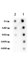
Dot Blot of Rabbit anti-Streptavidin Biotin Conjugated. Antigen: Lane 1 - Biotin conjugated Streptavidin Antibody. Lane 2 - Streptavidin Antibody. Load: 3-fold serial dilution starting at 200 ng. Secondary antibody: HRP Streptavidin at 1:40,000 for 60 min at RT. Block: 1% BSA-TTBS 30 min at RT.
Western Blot of Rabbit Anti-Trypsin Inhibitor Antibody and Rabbit Anti-Streptavidin Antibody. Lane 1: Trypsin Inhibitor reduced. Lane 2: Trypsin Inhibitor non-reduced. Lane 3: Streptavidin reduced. Lane 4: Streptavidin non-reduced. Load: ~1ug per lane. Primary antibody: Primary antibody at 1:1000 for overnight at 4°C. Secondary antibody: Dylight 649 conjugated Donkey anti rabbit secondary antibody at 1:10,000 for 45 min at RT. Block: MB-070 overnight at 4°C. Predicted/Observed size: 18.8 kDa, ~15 kDa for Streptavidin, 24 kDa, ~20 kDa for Trypsin Inhibitor. Other band(s): none.
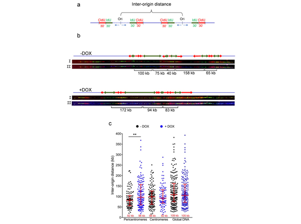
Origin of replication density in shTERF2-expressing HeLa cells. (a) Scheme showing the experimental setting used to measure the distance between two origins of replication (inter-origin distance; IOD); (b) Representative images of DNA combing at pericentromeres showing only the signal of IdU (green), IdU (red) top image (I) or with satellite III PNA probe signal (blue) bottom image (II). Green and red arrows indicate IdU and CldU labeling, respectively. IOD distance is shown below the images; (c) Quantification of IOD expressed in kilobases (kb) for HeLa cells expressing a doxycycline (DOX)-inducible shTERF2 system. Median length is shown at the bottom of the scatter plots. Bars represent the median +/- interquartile range (**?p?< 0.001; Mann?Whitney U test). Fig 2. PMID:?33804994
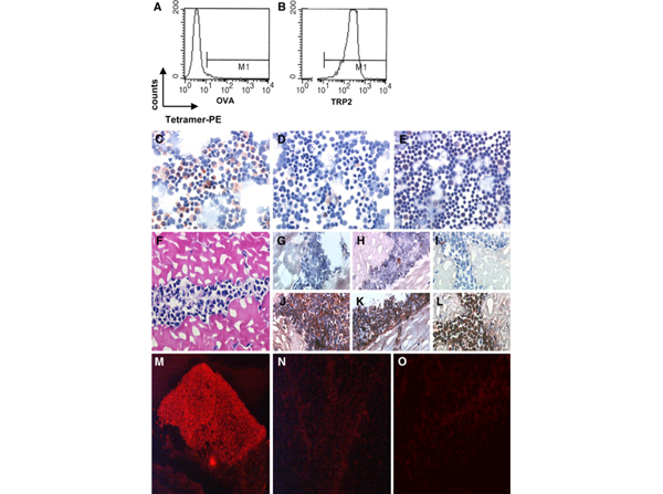
In situ detection of antigen-specific T cells. Flow cytometric analysis of tetramer binding by LP9 T cells, control tetramer H-2Kb-OVA (a) and relevant tetramer H-2Kb-TRP2180?(b). Positive staining of a cytological preparation containing LP9 cells and normal C57BL splenocytes was only observed with H-2Kb-TRP2180?tetramer (c) and not without tetramer (negative control) (d). Staining of normal splenocytes with H-2Kb-TRP2180?tetramer did not result in a positive signal (e). H&E-stained section of LP9-containing muscle tissue (f). Improved morphology and signal to noise ratios of in situ detection of antigen specific T cells in LP9-containing muscle tissue sections stained histochemically with specific tetramers (j?l) or negative control solution (without tetramer) (g?i) using the ABC-peroxidase kit with AEC (red-brown staining) as substrate.?g,?j?Without blocking with normal goat serum and PFA-fixation prior to staining.?h,?k?With normal goat serum blocking but without PFA-fixation prior to staining.?i,?l?With blocking with normal goat serum and PFA-fixation prior to staining. LP9-containing muscle tissue sections stained with or without tetramers with fluorescent microscopy visualization. Positive staining was observed after incubation with specific H-2Kb-TRP2180?tetramers (m) and not without the addition of tetramer (n) or after incubation with control tetramer H-2Kb-OVA. Fig 1.PMID:?17440724
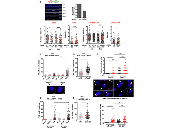
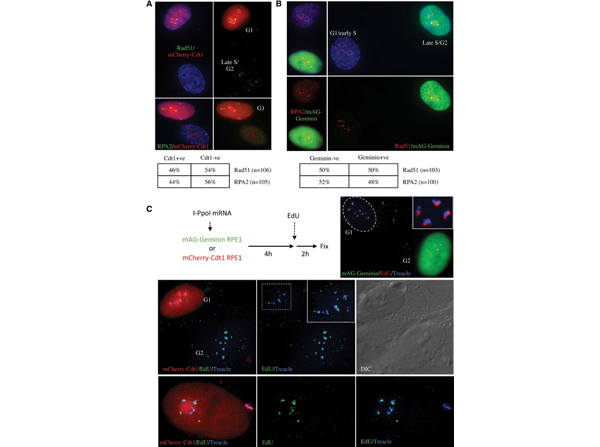
HR factors are recruited, and damage-induced DNA synthesis can be observed at I-PpoI-induced nucleolar caps in G1 cells. (A) I-PpoI transfection of RPE1 cells stably expressing FUCCI component mCherry-Cdt1 reveals that HR factors Rad51 and RPA2 are recruited comparably to nucleolar caps in G1 (red) and late S/G2 (nonred) cells. Quantification is shown?below. (B) I-PpoI transfection of RPE1 cells stably expressing FUCCI component mAG-Geminin reveals that HR factors Rad51 and RPA2 are recruited comparably to nucleolar caps in G1 (nongreen) and late S/G2 (green) cells. Quantification is shown?below. (C) RPE1 cells stably expressing FUCCI component mAG-Geminin or mCherry-Cdt1 were transfected with I-PpoI mRNA; 4 h after transfection, fresh medium containing EdU was added to cells. Following a 2-h pulse of EdU labeling, cells were fixed. Treacle antibodies detected damage-induced nucleolar caps. Damage-induced DNA synthesis was visualized using biotin-azide and fluorophore-conjugated streptavidin (see the Materials and Methods for details). In the?top right?panel, EdU incorporation can be observed at damage-induced nucleolar caps in G1 (nongreen) mAG-Geminin cells. In the?middle?panels, EdU incorporation is observed in nucleolar caps in both G1 (red) and G2 (nonred) mCherry-Cdt1 cells. A DIC image is shown at the?right. In the?bottom?panels, a single enlarged G1 cell from the same experiment is shown. Fig 6. PMID:?26019174
|
|
|
|
Dot Blot of Rabbit anti-Streptavidin Biotin Conjugated. Antigen: Lane 1 - Biotin conjugated Streptavidin Antibody. Lane 2 - Streptavidin Antibody. Load: 3-fold serial dilution starting at 200 ng. Secondary antibody: HRP Streptavidin at 1:40,000 for 60 min at RT. Block: 1% BSA-TTBS 30 min at RT.
|
|
| 別品名 |
rabbit anti-Streptavidin Antibody biotin Conjugation, biotin conjugated rabbit anti-Streptavidin antibody, Anti-Streptavidin BAC Antibody
|
| 交差種 |
Streptomyces avidinii
|
| 適用 |
Dot Blot
|
| 免疫動物 |
Rabbit
|
| 標識物 |
Biotin
|
| 精製度 |
Ig fraction - Ion Exchange /Gel Filtration
|
| 参考文献 |
[[Cosmo Reference Collection]][Pub Med ID]17440724,18951180,23811686,26019174,35877881
|
|
| メーカー |
品番 |
包装 |
|
RKL
|
200-4695
|
10 MG
|
※表示価格について
| 当社在庫 |
なし
|
| 納期目安 |
約10日
|
| 法規制 |
毒
|
| 保存温度 |
4℃
|
|
※当社では商品情報の適切な管理に努めておりますが、表示される法規制情報は最新でない可能性があります。
また法規制情報の表示が無いものは、必ずしも法規制に非該当であることを示すものではありません。
商品のお届け前に最新の製品法規制情報をお求めの際はこちらへお問い合わせください。
|
※当社取り扱いの試薬・機器製品および受託サービス・創薬支援サービス(納品物、解析データ等)は、研究用としてのみ販売しております。
人や動物の医療用・臨床診断用・食品用としては、使用しないように、十分ご注意ください。
法規制欄に体外診断用医薬品と記載のものは除きます。
|
|
※リンク先での文献等のダウンロードに際しましては、掲載元の規約遵守をお願いします。
|
|
※CAS Registry Numbers have not been verified by CAS and may be inaccurate.
|


