

| 別品名 |
FITC
|
| 種由来 |
Escherichia coli
|
| 標識物 |
Fluorescein Isothiocyanate
|
| 精製度 |
Ig fraction - Ion Exchange /Gel Filtration
|
| 適用 |
Western Blot
|
| 免疫動物 |
Rabbit
|
| Accession No.(Gene/Protein) |
NP_414878, P00722
|
| Tag情報 |
b-GAL
|
| 形状 |
凍結乾燥品
|
| [注意事項] |
濃度はロットによって異なる可能性があります。メーカーDS及びCoAからご確認ください。
|
|
※サムネイル画像をクリックすると拡大画像が表示されます。
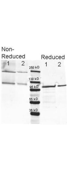
Western blotting using Rockland's anti b Galactosidase antibody. Lane 1 shows 80 ng and lane 2 shows 20 ng loaded onto gel. Results for non reducing conditions of SDS PAGE prior to transfer to nitrocellulose are shown on the left side of the figure; results obtainined under reducing conditions are shown on the right. Blots were blocked overnight at 4 C with Blocking Buffer for Fluorescent Western Blotting (p/n MB 070). The membrane was probed with anti b Galactosidase diluted to 1:10,000. Reaction occurred overnight at 4C. Dylight649TM conjugated Gt a anti Rabbit IgG (p/n 611 143 120) was used for detection. Molecular weight estimation was made by comparison to a prestained MW marker (center).in lane M. Fluorescence image was captured using the VersaDocR Imaging System developed by BIO RAD. Other detection systems will yield similar results.
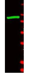
Western blot using Rockland's anti-b-Galactosidase antibody shows detection of a band at ~117 kDa (lane 1) corresponding to b-Gal present in a partially purified preparation (arrowhead). Approximately 1μg of protein was resolved on a 4-20% Tris-Glycine gel by SDS-PAGE and transferred onto nitrocellulose. After blocking, the membrane was probed with the primary antibody diluted to 1:1,000. Reaction occurred overnight at 4° C followed by washes and reaction with a 1:10,000 dilution of IRDyeR 800 conjugated Gt-a-Rabbit IgG (H&L) MX10 (611-132-122) for 45 min at room temperature (800 nm channel, green). Molecular weight estimation was made by comparison to prestained MW markers in lane M (700 nm channel, red). IRDyeR 800 fluorescence image was captured using the OdysseyR Infrared Imaging System developed by LI-COR. IRDye is a trademark of LI-COR, Inc. Other detection systems will yield similar results.
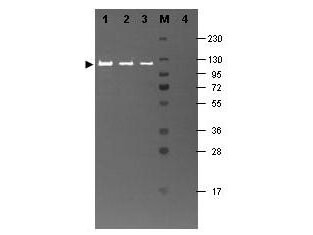
Western blotting using Rockland's Fluorescein conjugated anti-b-Galactosidase antibody shows a band at ~117 kDa (lanes 1 - 3) corresponding to 60 ng, 30 ng and 15 ng, respectively of b-Gal present in partially purified preparations (arrowhead). Lane 4 shows no cross reactivity with proteins present in a non-specific control E.coli lysate. Proteins were resolved on a 4-20% Tris-Glycine gel by SDS-PAGE and transferred to nitrocellulose and blocking using Blocking Buffer for Fluorescent Western Blotting (p/n MB-070). The membrane was probed with fluorescein conjugated anti-b-Galactosidase (p/n 200-4236) diluted to 1:10,000. Reaction occurred for 2 hours at room temperature. Molecular weight estimation was made by comparison to a prestained MW marker in lane M. Fluorescence image was captured using the VersaDocR Imaging System developed by BIO-RAD. Other detection systems will yield similar results.
|
|
|
|
Western blotting using Rockland's anti b Galactosidase antibody. Lane 1 shows 80 ng and lane 2 shows 20 ng loaded onto gel. Results for non reducing conditions of SDS PAGE prior to transfer to nitrocellulose are shown on the left side of the figure; results obtainined under reducing conditions are shown on the right. Blots were blocked overnight at 4 C with Blocking Buffer for Fluorescent Western Blotting (p/n MB 070). The membrane was probed with anti b Galactosidase diluted to 1:10,000. Reaction occurred overnight at 4C. Dylight649TM conjugated Gt a anti Rabbit IgG (p/n 611 143 120) was used for detection. Molecular weight estimation was made by comparison to a prestained MW marker (center).in lane M. Fluorescence image was captured using the VersaDocR Imaging System developed by BIO RAD. Other detection systems will yield similar results.
|
|
|
| メーカー |
品番 |
包装 |
|
RKL
|
200-4236
|
10 MG
|
※表示価格について
| 当社在庫 |
なし
|
| 納期目安 |
約10日
|
| 法規制 |
毒
|
| 保存温度 |
4℃
|
|

