|
※サムネイル画像をクリックすると拡大画像が表示されます。
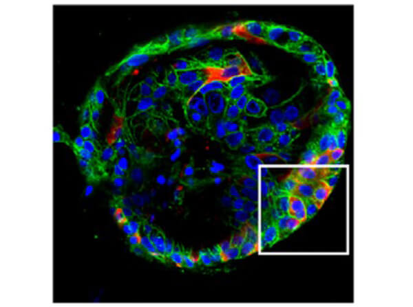
Immunofluorescence of Rabbit Anti-SARS-CoV Nucleocapsid (N) Antibody. Tissue: human Liver ductal organoids. Fixation: 4% PFA. Permeabilization: 0.25% Triton X-100. Antigen retrieval: not required. Primary antibody: Rabbit Anti-SARS-CoV (N) Antibody and Mouse Anti-E-Cadhedrin Antibody at 1:500 overnight at 2-8°C. Secondary antibody: Donkey Anti-Rabbit IgG CY3 Conjugated; Donkey Anti-Mouse IgG AlexaFluor 488 Conjugated for 1hr at RT. Nuclear Counterstain: DAPI. Staining showing Mock and Infected tissue: SARS-CoV Red signal, E-Cadhedrin green signal, with DAPI (blue) nuclear counterstain. [Zhao et al. (2020)]
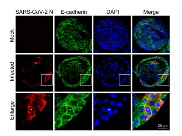
Immunofluorescence of Rabbit Anti-SARS-CoV Nucleocapsid (N) Antibody. Tissue: human Liver ductal organoids. Fixation: 4% PFA. Permeabilization: 0.25% Triton X-100. Antigen retrieval: not required. Primary antibody: Rabbit Anti-SARS-CoV (N) Antibody and Mouse Anti-E-Cadhedrin Antibody at 1:500 overnight at 2-8°C. Secondary antibody: Donkey Anti-Rabbit IgG CY3 Conjugated; Donkey Anti-Mouse IgG AlexaFluor 488 Conjugated for 1hr at RT. Nuclear Counterstain: DAPI. Staining of Infected and Merged: SARS-CoV Red signal, E-Cadhedrin green signal, with DAPI (blue) nuclear counterstain. [Zhao et al. (2020)]
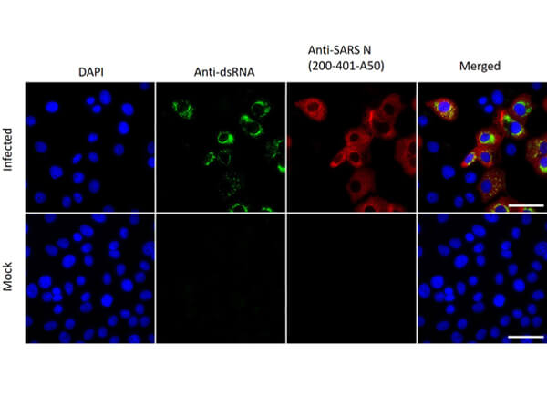
Immunofluorescence assay using Rabbit Anti-SARS-CoV Nucleocapsid (N) Antibody, showing viral protein synthesis. VeroE6 cells were infected with rSARS-CoV-2. Mock: non-infected cells. At 48 h.p.i, cells were fixed and prepared for immunofluorescence staining with primary antibodies directed against double-stranded RNA (dsRNA) [green] and SARS-CoV Nucleocapsid (N) [red]. Nuclear Counterstain DAPI [blue].
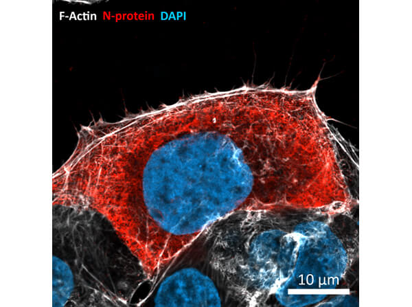
Immunofluorescence of Rabbit Anti-SARS CoV (M) Protein Antibody. Cells: Caco-2 cells 24 hours post-infection. Fixation: 4% PFA. Permeabilization: 0.3% Triton X-100. Blocking: 5% fetal calf serum/PBS. Primary Antibody: Anti-SARS-CoV nucleocapsid (N) protein at 1:1000 for 1 hour at RT. Staining: N-Protein [Red], F-actin [White], DAPI, [Blue]. Imaged: Zeiss LSM800 microscope. [Images Courtesy of AG Robert Grosse, Institute of Experimental and Clinical Pharmacology I, University of Freiburg/Svenja Ulferts and AG Georg Kochs Labs/Sebastian Weigang]. [Bouhaddou M et al. 2020]
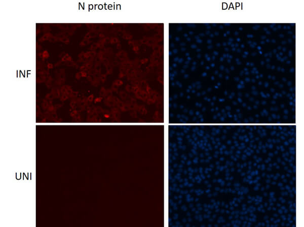
Immunofluorescence assay using Rabbit Anti-SARS-CoV Nucleocapsid (N) Antibody, showing viral protein detection. Vero E6 cells were either infected with the SARS-CoV-2 Washington isolate (INF) at an MOI of 0.1 or uninfected (UNI) for 24 hours. The cells were then fixed in 4% PFA and stained overnight at 4oC with primary antibodies directed against SARS-CoV Nucleocapsid (N) at 1:1000 dilution. Imaged using an anti-rabbit secondary conjugated to AlexaFluor 568 [red] and Nuclear Counterstain DAPI [blue]. Image Courtesy of Mohsan Saeed Lab/Da-Yuan Chen, National Emerging Infectious Diseases Laboratories (NEIDL), Boston University.
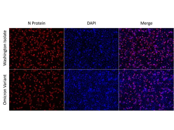
Immunofluorescence assay using Rabbit Anti-SARS-CoV Nucleocapsid (N) Antibody, showing viral protein detection. A549 cells over-expressing ACE2 were either infected with the SARS-CoV-2 Washington isolate or Omicron Variant at an MOI of 0.5 for 24 hours. The cells were then fixed in 10% Formalin and stained overnight at 4oC with primary antibodies directed against SARS-CoV Nucleocapsid (N) (Rockland p/n 200-401-A50) at 1:1000 dilution. Imaged using an anti-rabbit secondary conjugated to AlexaFluor 568 [red] and Nuclear Counterstain DAPI [blue]. Image Courtesy of Mohsan Saeed Lab/Da-Yuan Chen, National Emerging Infectious Diseases Laboratories (NEIDL), Boston University.
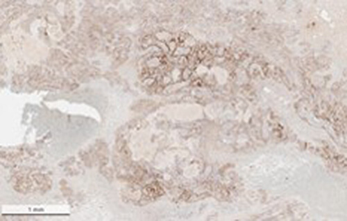
Immunohistochemistry results showing positive staining in placenta tissue using Anti-SARS-CoV Nucleocapsid (N) Antibody.
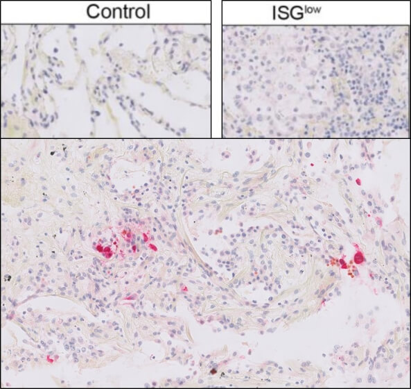
Immunohistochemistry of Rabbit Anti-SARS CoV Nucleocapsid Antibody. Pre-treated tissue: (A) human lung control, (B) interferon stimulated genes (ISG) low, (C) ISG high human COVID-19 lung: interferon stimulated genes (ISGs). [One pattern showes high expression of interferon stimulated genes (ISGs) and cytokines, high viral loads and limited pulmonary damage, the other pattern showes severely damaged lungs, low ISGs, low viral loads and abundant immune infiltrates.] Primary Antibody: Anti-SARS-CoV at 1:6400. Staining and Detection with Bond Polymer Refine Red Detection. [Courtesy of Mertz K. Profiles of COVID-19 lungs.]
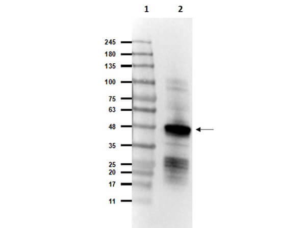
Western Blot of Rabbit Anti-SARS CoV Nucleocapsid (N) Protein Antibody. Lane 1: Opal Prestained Molecular Weight Marker (p/n MB-210-0500). Lane 2: SARS CoV Nucleocapsid (N) Protein [20ng]. Primary Antibody: Anti-SARS CoV Nucleocapsid (N) Protein Antibody at 1.0μg/mL overnight at 2-8°C. Secondary Antibody: Goat Anti-Rabbit IgG HRP (p/n 611-1302) at 1:40000 for 30mins at RT. Block: BlockOut Buffer (p/n MB-073). Predicted MW: ~46kDa. Observed MW: ~48kDa.
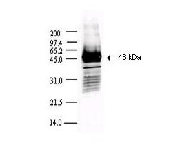
Western blot using Rockland's Protein A Purified anti-SARS CoV Nucleocapsid (N) protein antibody shows detection of a 46-kDa band corresponding to the protein. Approx. 100 ng of protein was loaded for SDS-PAGE and transferred onto nitrocellulose. The blot was incubated with a 1:5,000 dilution of the antibody at room temperature for 1 h followed by detection using IRDye?800 labeled Goat-a-Rabbit IgG [H&L] (611-132-122) diluted 1:10,000. The fluorescence image was captured using the OdysseyR Infrared Imaging System developed by LI-COR. IRDye is a trademark of LI-COR, Inc. Other detection systems will yield similar results.
|
|
|
|
Immunofluorescence of Rabbit Anti-SARS-CoV Nucleocapsid (N) Antibody. Tissue: human Liver ductal organoids. Fixation: 4% PFA. Permeabilization: 0.25% Triton X-100. Antigen retrieval: not required. Primary antibody: Rabbit Anti-SARS-CoV (N) Antibody and Mouse Anti-E-Cadhedrin Antibody at 1:500 overnight at 2-8°C. Secondary antibody: Donkey Anti-Rabbit IgG CY3 Conjugated; Donkey Anti-Mouse IgG AlexaFluor 488 Conjugated for 1hr at RT. Nuclear Counterstain: DAPI. Staining showing Mock and Infected tissue: SARS-CoV Red signal, E-Cadhedrin green signal, with DAPI (blue) nuclear counterstain. [Zhao et al. (2020)]
|
|
| 別品名 |
rabbit anti-Sars Nucleocapsid Protein Antibody, rabbit anti-Sars-CoV Nucleocapsid (N) Protein Antibody, N antibody, N structural protein antibody, NC antibody, Nucleocapsid protein antibody, Nucleoprotein antibody, SARS coronavirus N protein antibody, SARS CoV antibody, SARSCoV antibody, Severe acute respiratory syndrome antibody, A50 Antibody, A50 SARS, COVID
|
| 交差種 |
Virus
|
| 適用 |
Western Blot
Enzyme Linked Immunosorbent Assay
|
| 免疫動物 |
Rabbit
|
| 標識物 |
Unlabeled
|
| 精製度 |
Affinity Purified
|
| GENE ID |
1489678
|
| Accession No.(Gene/Protein) |
30173007, P59595
|
| 参考文献 |
[Pub Med ID]33622961
|
| [注意事項] |
濃度はロットによって異なる可能性があります。メーカーDS及びCoAからご確認ください。
|
|
| メーカー |
品番 |
包装 |
|
RKL
|
200-401-A50
|
500 UG
|
※表示価格について
| 当社在庫 |
なし
|
| 納期目安 |
約10日
|
| 法規制 |
毒
|
| 保存温度 |
4℃
|
|
※当社では商品情報の適切な管理に努めておりますが、表示される法規制情報は最新でない可能性があります。
また法規制情報の表示が無いものは、必ずしも法規制に非該当であることを示すものではありません。
商品のお届け前に最新の製品法規制情報をお求めの際はこちらへお問い合わせください。
|
※当社取り扱いの試薬・機器製品および受託サービス・創薬支援サービス(納品物、解析データ等)は、研究用としてのみ販売しております。
人や動物の医療用・臨床診断用・食品用としては、使用しないように、十分ご注意ください。
法規制欄に体外診断用医薬品と記載のものは除きます。
|
|
※リンク先での文献等のダウンロードに際しましては、掲載元の規約遵守をお願いします。
|
|
※CAS Registry Numbers have not been verified by CAS and may be inaccurate.
|
