|
※サムネイル画像をクリックすると拡大画像が表示されます。
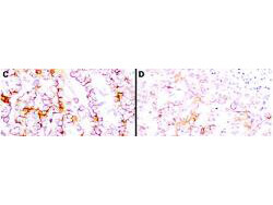
Immunohistochemistry using Rockland's anti-mesothelin antibody to react with two epitopes on mesothelin in PEFF human mesothelioma tissue sections treated by antigen retrieval methods. Anti-mesothelin primary antibodies were used at 10 μg/mL to label these sections as follows: C, MAb MB; and D, MAb MN followed by goat anti-mouse IgG conjugated to horseradish peroxidase at 25 μg/mL in 1% BSA/PBS for 30 minutes. (magnification, ×200; bar, 50 μm). Reprinted with permission from Clin.Cancer Res. 11(16):5840-6.
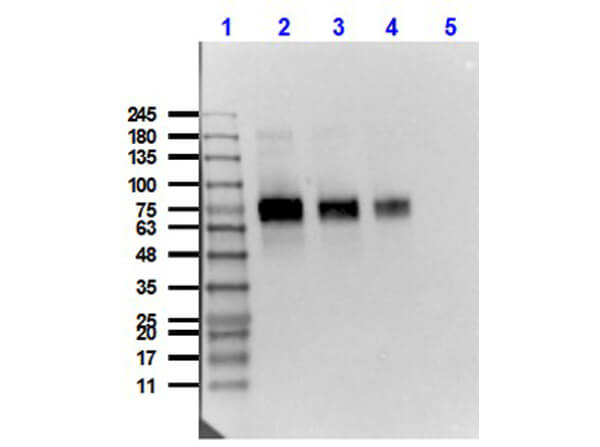
Western Blot of Mouse Anti-Mesothelin Antibody. Lane 1: Opal Prestained Molecular Weight Marker (p/n MB-210-0500). Lane 2: HeLa [10μg] + Mesothelin-Fc [0.1μg]. Lane 3: HeLa [10μg] + Mesothelin-Fc [0.05μg]. Lane 4: HeLa [10μg] + Mesothelin-Fc [0.02μg]. Lane 5: HeLa Whole Cell Lysate (p/n W09-000-364). Primary Antibody: Anti-Mesothelin at 1μg/mL overnight at 2-8°C. Secondary Antibody: Rabbit Anti-Mouse IgG HRP conjugated (p/n 610-4302) at 1:40,000 for 30 mins at RT. Block: BlockOut Buffer (p/n MB-073) 30 mins at RT. Exposure: 15 sec. Predicted MW: 40kDa Mesothelin + Fc region 30kDa. Observed MW: ~70-75kDa.
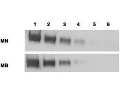
Western blotting using Rockland's anti-mesothelin antibodies to detect mesothelin-Fc at 100 ng (lane 1), 25 ng (lane 2), 6 ng (lane 3), 2 ng (lane 4) and 0.4 ng (lane 5). Lane 6 contains 50 ng of CDC25-Fc. Proteins were separated on 4-20% gradient gel by SDS-PAGE followed by transfer to PVDF membrane. Primary antibody was used at 1 μg/ml followed by reaction with ALP goat anti-mouse IgG and BCIP/NBT substrate. Reprinted with permission from Clin.Cancer Res. 11(16):5840-6.
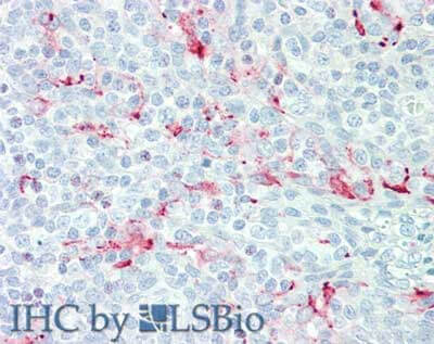
Immunohistochemistry of Mouse anti-Mesothelin antibody. Tissue: human tonsil. Fixation: formalin fixed paraffin embedded. Antigen retrieval: not required. Primary antibody: anti-Mesothelin antibody at 15 μg/mL for 1 h at RT. Secondary antibody: Peroxidase mouse secondary antibody at 1:10,000 for 45 min at RT. Staining: Mesothelin as precipitated red signal with hematoxylin purple nuclear counterstain.
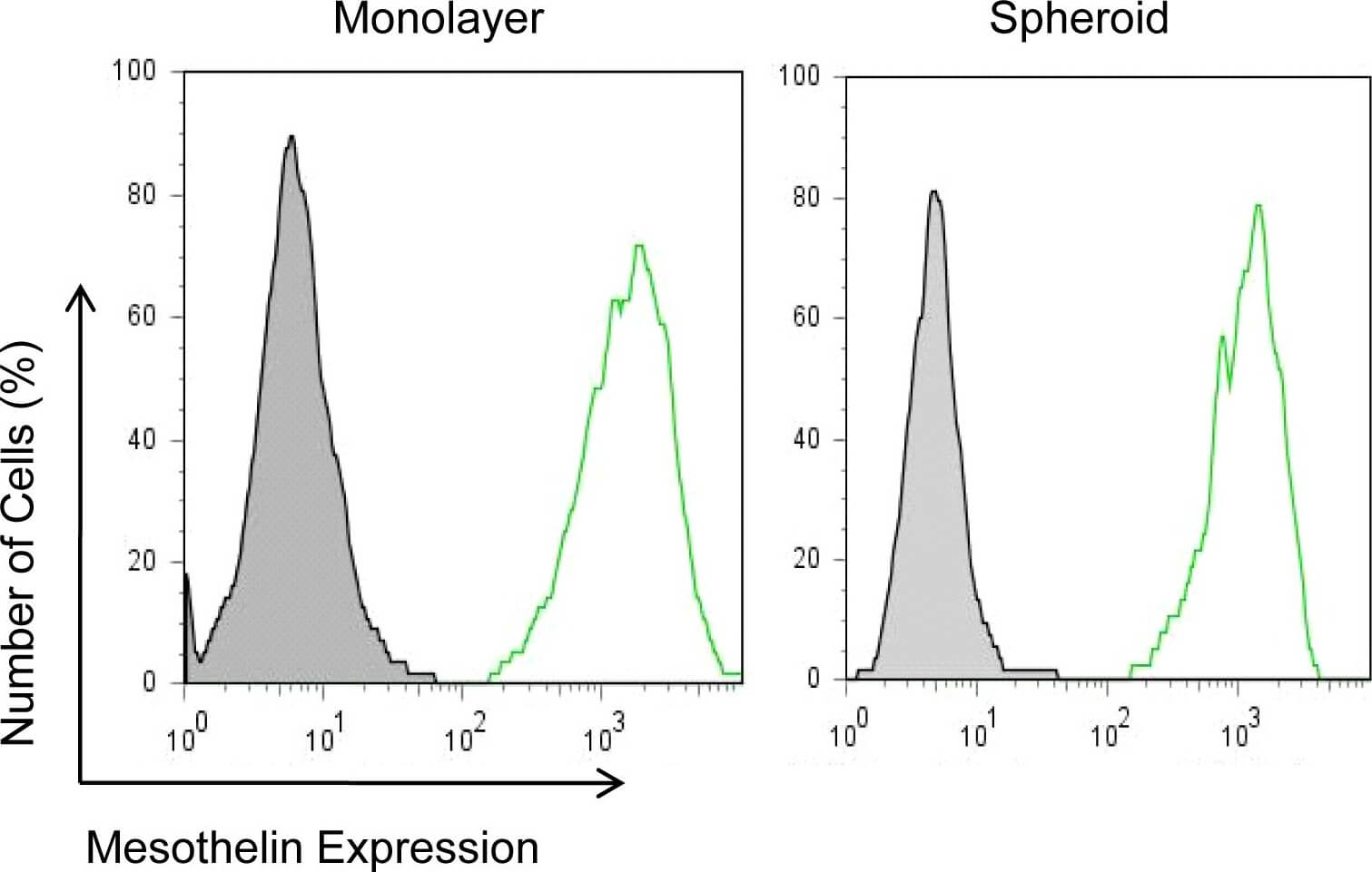
Mesothelin expression in mesothelioma monolayers and spheroids.NCI-H226 cells incubated with an anti-mesothelin mAb (MN) and detected with goat anti-mouse IgG conjugated with Alexa488 by flow cytometry. Figure provided by CiteAb. Source: PLoS One, PMID: 21305058.
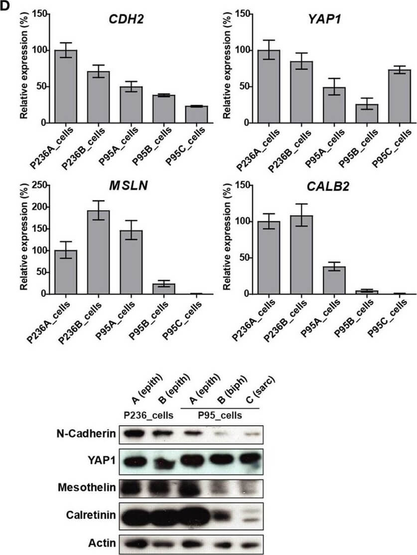
(A) Tumor processing flow-chart of 235 samples, which were processed for RNA extraction, cell culture and embedding either in PFA or OCT. Samples showing attributes which are grayed in the chart, were not used in the live cell biobank. (B) Circos whole genome copy number variations (CNVs) view of tumor and primary cell culture in patients malignant pleural mesothelioma (MPM) 236 and MPM95. The quilt plot highlights the CNV and SNVs in genes that are part of MPM landscape (2). (C) Immunofluorescence analysis of selected markers in primary culture from patient MPM236. Scale bar 200?μm. (D) Selected genes expression analysis at mRNA (upper panel) and protein (lower panel) level in primary cultures derived from samples from two patients, MPM236 and MPM95. The latter one underwent EMT during disease progression. (E) Selected genes expression analysis at mRNA in tumor samples from patients MPM236 and MPM95. (F) Significant correlation between gene expression changes in tumor and primary culture from patient MPM236 at passage 3. Figure provided by CiteAb. Source: Front Oncol, PMID: 29527515.
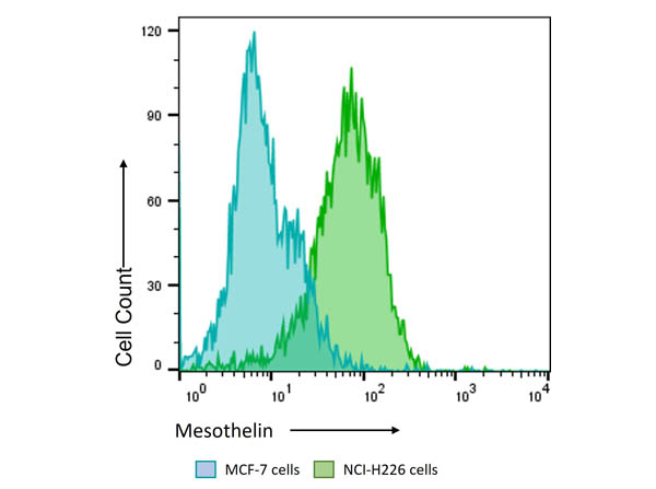
Flow Cytometry Results of Anti-Mesothelin (MOUSE) Monoclonal Antibody. The green histogram shows NCI-H226 cells and blue histogram shows MCF-7 cells. Both cell lines are stained with a 1:800 dilution Anti-Mesothelin (MOUSE) Monoclonal Antibody. The secondary antibody use was Anti-Mouse IgG (H&L) (GOAT) Antibody DyLight? 488 Conjugated (p/n 610-141-002, lot#43322) at the 1:400 dilution.
|
|
|
|
Immunohistochemistry using Rockland's anti-mesothelin antibody to react with two epitopes on mesothelin in PEFF human mesothelioma tissue sections treated by antigen retrieval methods. Anti-mesothelin primary antibodies were used at 10 μg/mL to label these sections as follows: C, MAb MB; and D, MAb MN followed by goat anti-mouse IgG conjugated to horseradish peroxidase at 25 μg/mL in 1% BSA/PBS for 30 minutes. (magnification, ×200; bar, 50 μm). Reprinted with permission from Clin.Cancer Res. 11(16):5840-6.
|
|
| 別品名 |
mouse anti-Mesothelin Antibody, Mesothelian, MN, MB, Pre-pro-megakaryocyte-potentiating factor, CAK1 antigen
|
| 交差種 |
Human
|
| 適用 |
Western Blot
Immunohistochemistry
Flow Cytometry
|
| 免疫動物 |
Mouse
|
| クローン |
MN-1
|
| 抗体クラス |
IgG2a
|
| 抗原部位 |
Extracellular domain
|
| 標識物 |
Unlabeled
|
| 精製度 |
Ig fraction - Protein A
|
| GENE ID |
10232
|
| Accession No.(Gene/Protein) |
53988378, Q13421
|
| Gene Symbol |
MSLN
|
| 参考文献 |
[Pub Med ID]32194667
|
| [注意事項] |
濃度はロットによって異なる可能性があります。メーカーDS及びCoAからご確認ください。
|
|
| メーカー |
品番 |
包装 |
|
RKL
|
200-301-A88S
|
25 UL
|
※表示価格について
| 当社在庫 |
なし
|
| 納期目安 |
約10日
|
| 保存温度 |
-20℃
|
|
※当社では商品情報の適切な管理に努めておりますが、表示される法規制情報は最新でない可能性があります。
また法規制情報の表示が無いものは、必ずしも法規制に非該当であることを示すものではありません。
商品のお届け前に最新の製品法規制情報をお求めの際はこちらへお問い合わせください。
|
※当社取り扱いの試薬・機器製品および受託サービス・創薬支援サービス(納品物、解析データ等)は、研究用としてのみ販売しております。
人や動物の医療用・臨床診断用・食品用としては、使用しないように、十分ご注意ください。
法規制欄に体外診断用医薬品と記載のものは除きます。
|
|
※リンク先での文献等のダウンロードに際しましては、掲載元の規約遵守をお願いします。
|
|
※CAS Registry Numbers have not been verified by CAS and may be inaccurate.
|
