
| 別品名 |
Cas like docking antibody, CASL antibody, Crk associated substrate related protein antibody, dJ49G10.2 antibody, dJ761I2.1 antibody, Enhancer of filamentation 1 antibody
|
| 抗原部位 |
a.a.82-398
|
| 種由来 |
Human
|
| 標識物 |
Unlabeled
|
| 精製度 |
Ig fraction - Protein A
|
| 適用 |
Western Blot
Immuno Fluorescence
Immunoprecipitation
|
| 免疫動物 |
Mouse
|
| 抗体クラス |
IgG1κ
|
| クローン |
2G9
|
| 交差種 |
Human
Mouse
Rat
|
| GENE ID |
4739
|
| Accession No.(Gene/Protein) |
Q14511
|
| Gene Symbol |
NEDD9
|
| 形状 |
滅菌済み液状品
|
| 参考文献 |
Merrill,R.A., See,A.W., Wertheim,M.L. and Clagett-Dame,M. (2004) Crk-associated substrate (Cas) family member, NEDD9, is regulated in human neuroblastoma cells and in the embryonic hindbrain by all-trans retinoic acid. Dev. Dyn. 231 (3), 564-575. Zheng,M. and McKeown-Longo,P.J. (2002) Regulation of HEF1 expression and phosphorylation by TGF-beta 1 and cell adhesion. J. Biol. Chem. 277 (42), 39599-39608. Law,S.F., Estojak,J., Wang,B., Mysliwiec,T., Kruh,G. and Golemis,E.A. (1996) Human enhancer of filamentation 1, a novel p130cas-like docking protein, associates with focal adhesion kinase and induces pseudohyphal growth in Saccharomyces cerevisiae. Mol. Cell. Biol. 16 (7), 3327-3337. Pugacheva EN, Golemis EA. (2005) The focal adhesion scaffolding protein HEF1 regulates activation of the Aurora-A and Nek2 kinases at the centrosome. Nat Cell Biol. 10:937-46.
|
| [注意事項] |
濃度はロットによって異なる可能性があります。メーカーDS及びCoAからご確認ください。
|
|
※サムネイル画像をクリックすると拡大画像が表示されます。
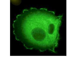
Immunofluorescence microscopy using Rockland's Monoclonal anti HEF1 antibody (clone 2G9) shows detection of HEF1 localized at focal adhesion sites. The antibody was used at a 1:500 dilution with a 3 sec exposure time. Personal Communication. Elena Pugacheva, Fox Chase Cancer Center, Philadelphia, PA.
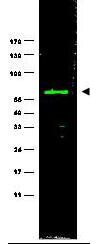
Western blot using Rockland's monoclonal anti-HEF1 antibody (clone 2G9) antibody shows detection of a ~92 kDa band corresponding to HEF1 in MCF7 lysate (p/n W09-000-360) [arrowhead]. Approximately 35 μg of lysate was loaded for SDS-PAGE followed by transfer onto nitrocellulose and reaction with a 1:1,000 dilution of anti-HEF1 antibody. Detection occurred using a 1:5,000 dilution of IRDyeR800 conjugated Sh-a-Mouse IgG [H&L] (p/n 610-632-002) for 45 min at room temperature (800 nm channel, green). Molecular weight estimation was made by comparison to prestained MW markers (indicated at left). IRDyeR800 fluorescence image was captured using the OdysseyR Infrared Imaging System developed by LI-COR. IRDye is a trademark of LI-COR, Inc. Other detection systems will yield similar results.
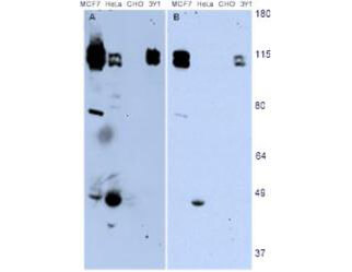
Western blotting using Rockland's monoclonal anti-HEF1 antibody (clone 2G9) shows detection of endogenous HEF1 present in various cell lines [MCF7, HeLa, CHO, 3Y1]. Panel A shows detection using a 15 min exposure. Panel B is the same blot exposed for 2 min. The doublet represents p105 and p115 staining. The lower MW band represents p55. 3Y1 cells are derived from rat fibroblast cells. Mouse 3T3 cells are also reactive (not shown). To date no staining has been noted on CHO cells. Personal Communication. Elena Pugacheva, Fox Chase Cancer Center, Philadelphia, PA.
|
|
|
|
Immunofluorescence microscopy using Rockland's Monoclonal anti HEF1 antibody (clone 2G9) shows detection of HEF1 localized at focal adhesion sites. The antibody was used at a 1:500 dilution with a 3 sec exposure time. Personal Communication. Elena Pugacheva, Fox Chase Cancer Center, Philadelphia, PA.
|
|
|
| メーカー |
品番 |
包装 |
|
RKL
|
200-301-904
|
100 UG
|
※表示価格について
| 当社在庫 |
なし
|
| 納期目安 |
約10日
|
| 保存温度 |
-20℃
|
|

