|
※サムネイル画像をクリックすると拡大画像が表示されます。
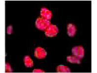
Anti ATM antibody showing overlay of anti-ATM pS1981 staining. Cells were fixed 15 min after 5 Gy (IR+) of irradiation, then labeled with antibody. See Kitagawa et al. for additional details
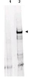
Detection of Monoclonal Anti-ATM with human derived HEK293 cell lysates treated with doxorubicin using Rockland's Protein A Purified Mab anti-ATM Protein Kinase pS1981 (clone 10H11.E12). A 370kDa band corresponding to phosphorylated ATM is detected (arrowhead, lane 2). The lysate was prepared with HALT phosphatase inhibitor (Pierce). Pre-incubation of antibody with immunizing phospho peptide negates specific staining (lane 1). Approximately 30μg of lysate was added to each lane of an SDS-PAGE gel under non-reducing conditions. The protein was transferred to nitrocellulose using standard methods. After blocking the membrane was probed with the primary antibody diluted 1:500 overnight at 4°C followed by washes and reaction with a 1:10,000 dilution of IRDye?800 conjugated Gt-a-Mouse IgG [H&L] (p/n 610-132-121) for 40 min at room temperature. LICOR's OdysseyR Infrared Imaging System was used to scan and process the image. Other detection systems will yield similar results.
Rockland Mouse Anti-ATM Protein Kinase pS1981 Antibody.
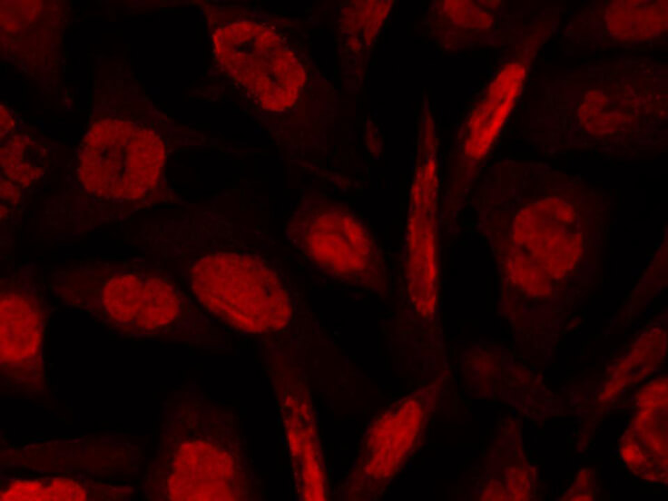
Rockland's anti-ATM pS1981 mouse monoclonal antibody (p/n 200-301-400) detects ATM phosphorylated on Ser 1981 by Indirect immunofluorescence microscopy. Shown are hTCEpi cells (courtesy of Dr. Danielle Robertson) infected with HSV-1 at MOI 5.0 and fixed at 8 hpi with 3% paraformaldehyde/2% sucrose for 10 min. After rinsing, cells were permeabilized with 0.5% Triton X-100 for 5 min, blocked with 3% BSA for 30 min, and stained with Rockland's primary anti-ATM pS1981 antibody overnight at 5μg/mL (1:200). Secondary staining was performed with Alexa Fluor 594 anti-mouse antibody. Images were taken with Olympus AX70 compound epifluorescence microscope equipped with Spot RT Slider camera. Experiment was performed by Oleg Alekseev in the laboratory of Dr. Jane Azizkhan-Clifford at Drexel University College of Medicine.
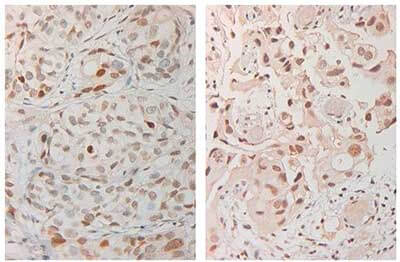
Immunohistochemistry with anti-ATM Antibody. Tissue: Human Bladder Cancer. Fixation: FFPE buffered formalin 10% conc. Ag Retrieval: HIER citrate buffer pH6 (left) or HIER EDTA pH9 (Right). Primary antibody: anti-ATM at 2 ug/ml for 2 hr. Secondary Ab: anti-rabbit polymer HRP for 20min at RT.
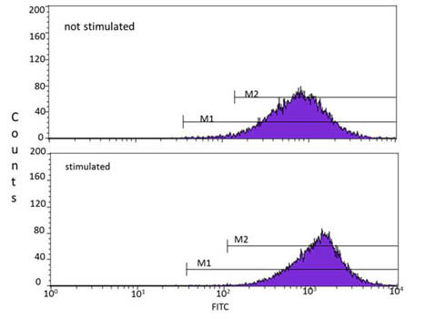
Flow Cytometry of Mouse anti-ATMpS1981 antibody. Cells: HEK293. Stimulation: none ? top image, 0.1mg/ml Zeocin for 3 hr ? bottom image. Primary antibody: anti-ATM pS1981 antibody at 5 μg/mL for 30 min at 4°C. Secondary antibody: anti-mouse IgG FITC at 1μg/ml, 30min at 4°C IN THE DARK.
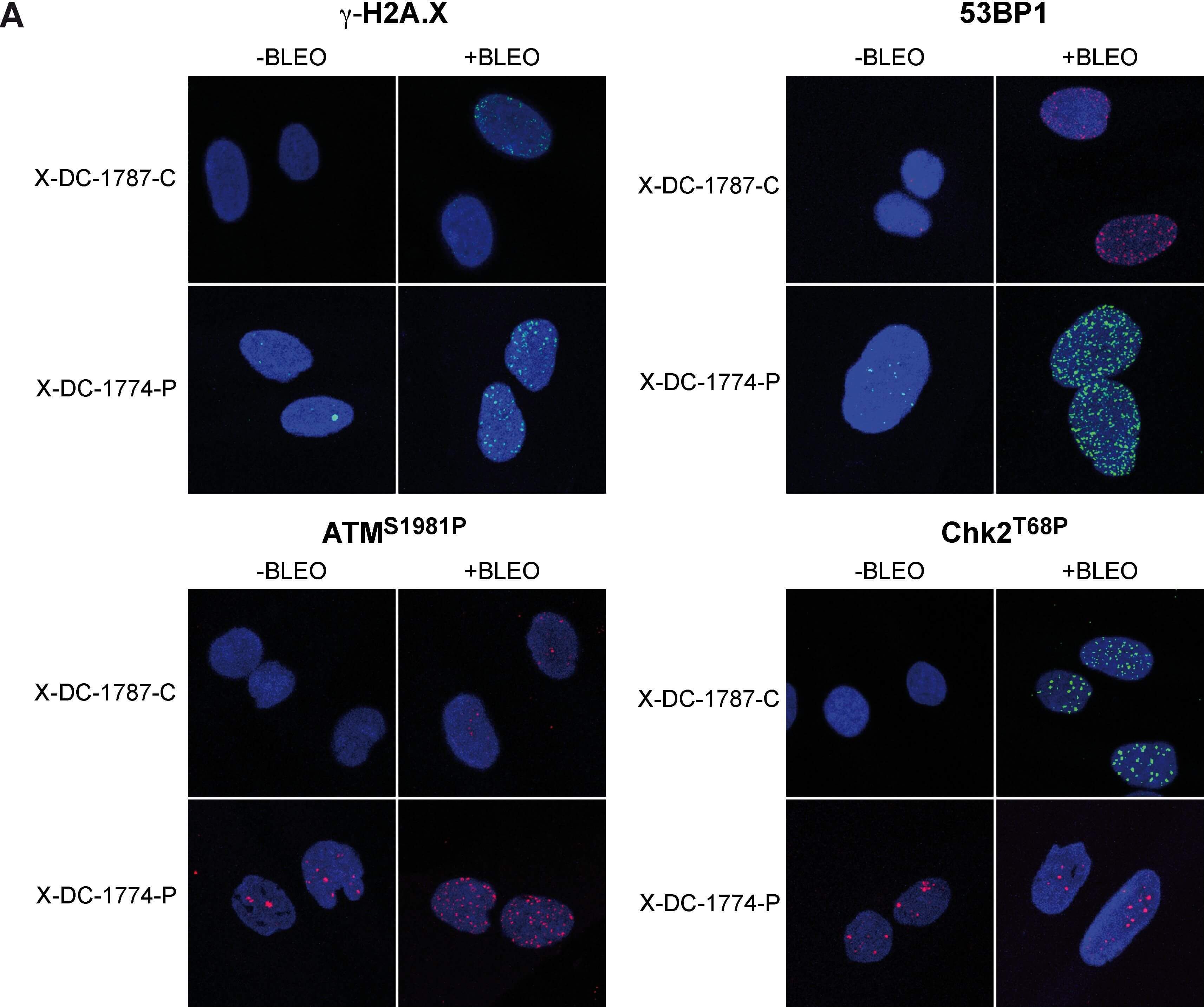
DNA damage signaling in X-DC patient cells.(A) Immunofluorescence staining of DNA damage proteins. Control X-DC-1787-C and patient X-DC-1774-P cells were, either not treated (-Bleo) or treated (+Bleo) with bleomycin (10 μg/ml) for 24 hours, fixed and incubated with antibodies against γ-H2AX, 53BP1, p-ATM or p-CHK1 and secondary fluorescent antibodies. Nuclear DNA was counterstained with DAPI (blue). (B). Quantification of γ-H2A.X foci, pATM, 53BP1 and pCHK2 associated foci in X-DC-1787-C and X-DC-1774-P cells. More than 200 cells were analyzed in each cell line and indicated as the average number of foci/cell. Asterisks indicate significant differences in relation to control cells lines or to untreated cells. Average values and standard deviations of two independent experiments are shown. Experiments were repeated 3 times with similar results. Figure provided by CiteAb. Source: PLoS One, PMID: 24987982.
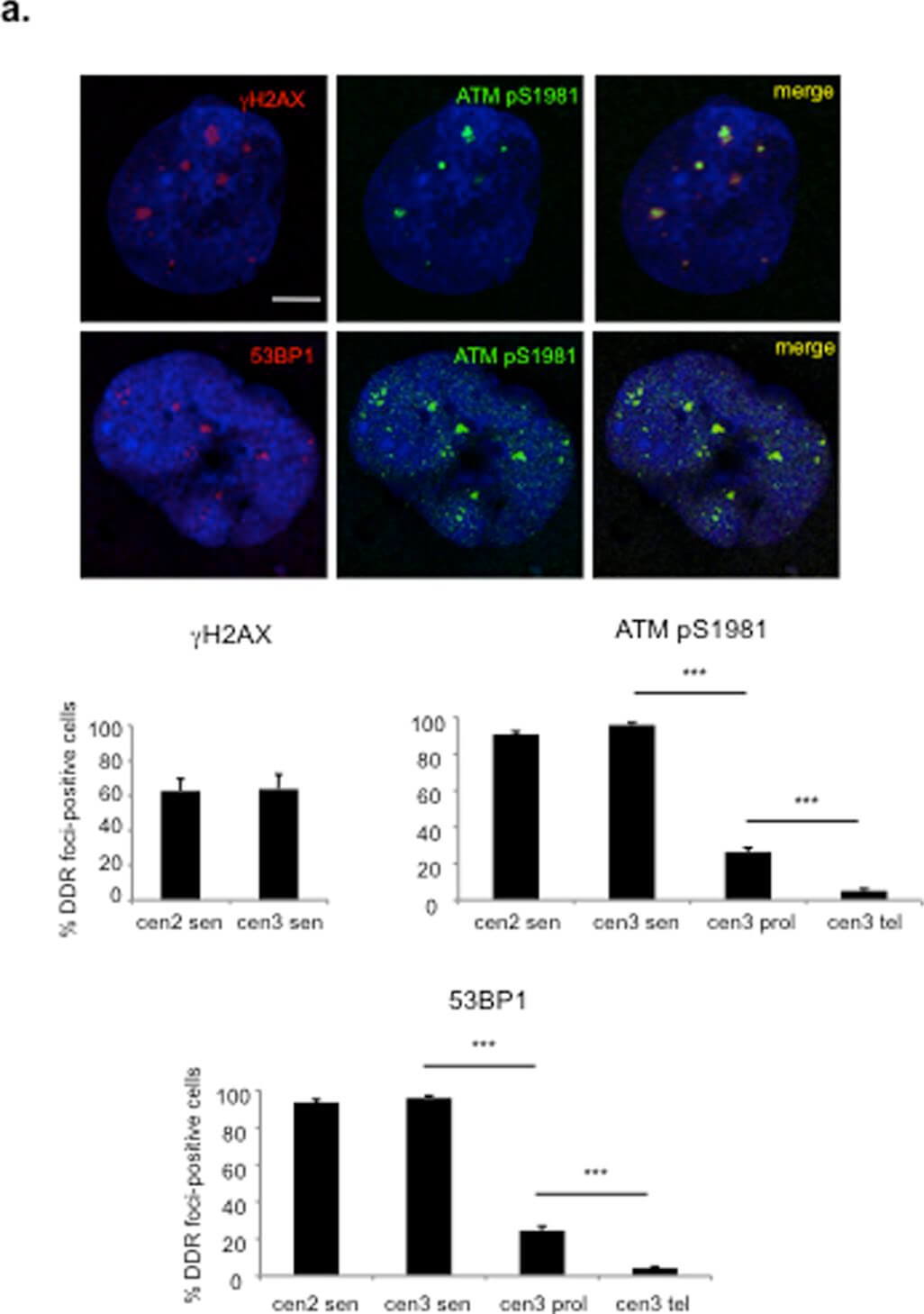
Telomere-initiated senescent cells retain active DDR foci for years after senescence establishment.a. DDR, in the form of ATM pS1981 foci co-localizing with 53BP1 and γH2AX foci, is detectable three years after senescence establishment. Scale bar, 10 μm. Below, bar graphs show the percentage of cells positive ± s.e.m. for the indicated DDR markers, in senescent (sen), early passage proliferating (prol) or telomerized proliferating (tel) skin fibroblasts from two independent centenarian donors (cen2 and cen3). Cells were considered positive if bearing more than 3 DDR foci (*** p-value <0.001). b. SA-β-gal staining of the two batches, cen2 and cen3, is shown together with the percentage of BrdU-positive cells. Scale bar, 100 μm. Figure provided by CiteAb. Source: PLoS One, PMID: 25340529.
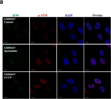
ATM is activated by spermidine in GM00637 cells.GM00637 cells with or without KU55933 pretreatment (a,b), and GM05849 cells (c) were exposed to 50?μM spermidine or CCCP, followed by immunofluorescence analyses of total and p-ATM on Ser-1981. The scale bar is 10?μm. Ratios of cells expressing p-ATM Ser-1981 to cells expressing total ATM were presented (d). 20?45 cells/condition from three experiments were collected. Values are mean?±?SD, *p?<?0.05 vs. control. Figure provided by CiteAb. Source: Sci Rep, PMID: 27089984.
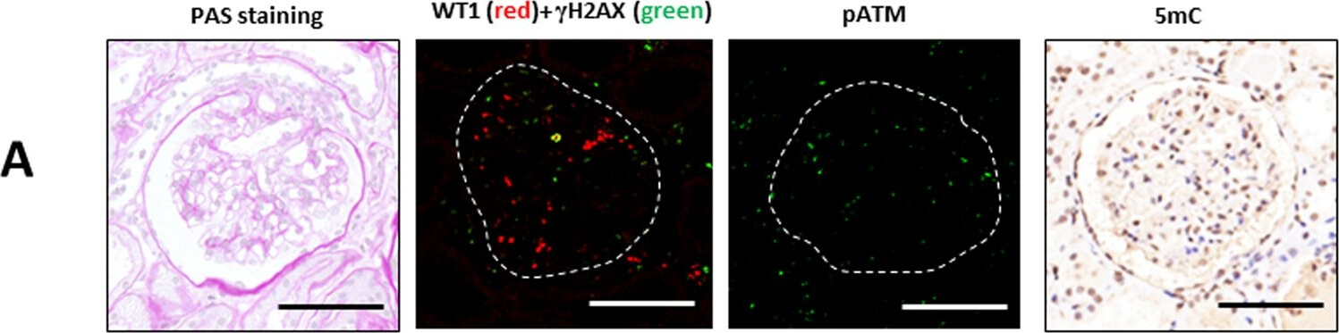
Immunostaining of γH2AX, WT1 and 5mC in patients with IgA nephropathy and controls. Examples of PAS staining and immunostaining with γH2AX (green) and WT1 (red), pATM and 5mC in glomeruli of IgA nephropathy and controls. (A) A control kidney sample of 44-year-old female. Scale bars: 50 μm. Figure provided by CiteAb. Source: Sci Rep, PMID: 31937846.
|
|
|
|
Anti ATM antibody showing overlay of anti-ATM pS1981 staining. Cells were fixed 15 min after 5 Gy (IR+) of irradiation, then labeled with antibody. See Kitagawa et al. for additional details
|
|
| 別品名 |
mouse anti-ATM antibody, mouse anti-ATMpS1981 antibody, mouse anti- ATM pS1981 antibody, DKFZp781A0353 antibody, Human phosphatidylinositol 3 kinase homolog antibody, MGC74674 antibody, Serine protein kinase ATM antibody, T cell prolymphocytic leukemia antibody
|
| 交差種 |
Human
Mouse
Rat
|
| 適用 |
Western Blot
Enzyme Linked Immunosorbent Assay
Immuno Fluorescence
Flow Cytometry
|
| 免疫動物 |
Mouse
|
| クローン |
10H11.E12
|
| 抗体クラス |
IgG1κ
|
| 抗原部位 |
a.a.1974-1988
|
| 標識物 |
Unlabeled
|
| 精製度 |
Ig fraction - Protein A
|
| 翻訳後修飾 |
リン酸化
|
| GENE ID |
472
|
| Accession No.(Gene/Protein) |
NP_000042.3, Q13315
|
| Gene Symbol |
ATM
|
| 参考文献 |
[Pub Med ID]15140942
|
| [注意事項] |
濃度はロットによって異なる可能性があります。メーカーDS及びCoAからご確認ください。
|
|
| メーカー |
品番 |
包装 |
|
RKL
|
200-301-400
|
100 UG
|
※表示価格について
| 当社在庫 |
なし
|
| 納期目安 |
約10日
|
| 保存温度 |
-20℃
|
|
※当社では商品情報の適切な管理に努めておりますが、表示される法規制情報は最新でない可能性があります。
また法規制情報の表示が無いものは、必ずしも法規制に非該当であることを示すものではありません。
商品のお届け前に最新の製品法規制情報をお求めの際はこちらへお問い合わせください。
|
※当社取り扱いの試薬・機器製品および受託サービス・創薬支援サービス(納品物、解析データ等)は、研究用としてのみ販売しております。
人や動物の医療用・臨床診断用・食品用としては、使用しないように、十分ご注意ください。
法規制欄に体外診断用医薬品と記載のものは除きます。
|
|
※リンク先での文献等のダウンロードに際しましては、掲載元の規約遵守をお願いします。
|
|
※CAS Registry Numbers have not been verified by CAS and may be inaccurate.
|
