
| 別品名 |
Structural maintenance of chromosomes protein 1B antibody, SMC1beta protein antibodySMC1B antibody, SMC1L2 antibody
|
| 抗原部位 |
a.a.951-962
|
| 種由来 |
Human
|
| 標識物 |
Unlabeled
|
| 精製度 |
Ig fraction - Protein G
|
| 適用 |
Western Blot
Enzyme Linked Immunosorbent Assay
Immunohistochemistry
|
| 免疫動物 |
Mouse
|
| 抗体クラス |
IgG1κ
|
| クローン |
5D11.G5
|
| 交差種 |
Human
Mouse
|
| 翻訳後修飾 |
リン酸化
|
| GENE ID |
27127
|
| Accession No.(Gene/Protein) |
Q8NDV3
|
| Gene Symbol |
SMC1B
|
| 形状 |
滅菌済み液状品
|
| 参考文献 |
[Pub Med ID]23587483, 15175241, 19450527, 16432227, 33627431
|
| [注意事項] |
濃度はロットによって異なる可能性があります。メーカーDS及びCoAからご確認ください。
|
|
※サムネイル画像をクリックすると拡大画像が表示されます。
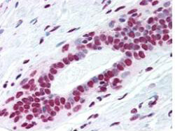
Rockland's Protein G Purified Mab anti SMC1 pS957 antibody was used at a 2.5 ug/ml to detect nuclear signal in a variety of tissues including multi human, multi brain and multi cancer slides. This image shows moderate to strong nuclear anti SMC1 pS957 staining of human breast ductal epithelium. Tissue was formalin fixed and paraffin embedded. The image shows localization of the antibody as the precipitated red signal, with a hematoxylin purple nuclear counterstain. Personal Communication, Tina Roush, LifeSpan Biosciences, Seattle, WA.
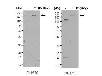
Western blot of gamma irradiated (+ lanes) and mock-irradiated (- lanes) human GM536 lymphoblastoid cell lysate (left panel) and mouse NIH3T3 cell lysate (right panel). Rockland's Protein G Purified Mab anti-SMC1 pS957 detects a 160 kDa band corresponding to phosphorylated SMC1. The antibody does not react with non-phosphorylated SMC1 present in the human control lane. Non specific binding may occur in control lanes of lysates from mouse cell origins. The cell lysates were prepared in a RIPA buffer containing 200 mM NaCl, and 20 ug protein was loaded per lane. A 4-12% Bis-Tris gradient gel (Invitrogen) was used for SDS-PAGE. The membrane was probed with the primary antibody at 10ug/ml for 1 h at 20C followed by washes and reaction with a 1:1000 dilution of HRP conjugated Dnky-a-Mouse IgG [H&L] (code 610-703-124) for 30 min.
|
|
|
|
Rockland's Protein G Purified Mab anti SMC1 pS957 antibody was used at a 2.5 ug/ml to detect nuclear signal in a variety of tissues including multi human, multi brain and multi cancer slides. This image shows moderate to strong nuclear anti SMC1 pS957 staining of human breast ductal epithelium. Tissue was formalin fixed and paraffin embedded. The image shows localization of the antibody as the precipitated red signal, with a hematoxylin purple nuclear counterstain. Personal Communication, Tina Roush, LifeSpan Biosciences, Seattle, WA.
|
|
|
| メーカー |
品番 |
包装 |
|
RKL
|
200-301-397
|
100 UG
|
※表示価格について
| 当社在庫 |
なし
|
| 納期目安 |
約10日
|
| 保存温度 |
-20℃
|
|

