|
※サムネイル画像をクリックすると拡大画像が表示されます。
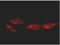
Immunofluorescence microscopy of HeLa cells using anti-p53. Rockland's Protein A purified Mab anti-p53 was used at a 1:100 dilution in 10% normal goat serum in PBS and reacted overnight at 4° C. After washes cells were incubated with a 1:500 dilution of AlexaFluor594 Goat-a-Mouse IgG diluted in normal goat serum for 1 h at room temperature. Personnel Communication. Kuldeep Patel, Loyola University.
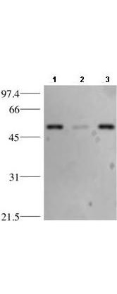
Western blotting using anti-p53 Antibody. Lane 1: HeLa whole cell lysate (p/n W09-000-364), Lane 2: HeLa cytosol fraction, and Lane 3: HeLa nuclear extract (p/n W09-001-367). Load of 15 μg was separated by 10% SDS-PAGE and transferred to nitrocellulose membrane. The membrane was blocked with 3% milk/TBST for 1 h at room temperature followed by incubation with Rockland's Protein A purified Mab anti-p53 antibody overnight at 4° C diluted 1:1,500 in blocking solution. The membrane was washed 3X with TBST and then incubated with a 1:2,000 dilution of HRP Goat-a-Mouse IgG (p/n 610-103-121) diluted in blocking buffer for 1 h at room temperature. After final washes the proteins reactive on the membrane were detected using ECL. Other detection systems will yield similar results. Personnel Communication. Kuldeep Patel, Loyola University.
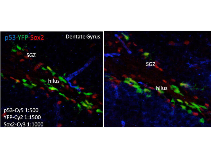
Immunofluorescence of Mouse anti-p53 antibody. Tissue: human brain. Fixation: free-floating. Antigen retrieval: not required. Primary antibody: anti-human-p53 antibody at 1:500 for 1 h at RT. Co-stained with YFP and Sox2 antibodies. Localization: p53 is nuclear and cytoplasmic. Staining: p53 as precipitated blue with Cy5, YFP as precipitated green with Cy2, and Sox2 as precipitated red with Cy3. z‐stacks from confocal expressed as one composite focal plain.
|
|
|
|
Immunofluorescence microscopy of HeLa cells using anti-p53. Rockland's Protein A purified Mab anti-p53 was used at a 1:100 dilution in 10% normal goat serum in PBS and reacted overnight at 4° C. After washes cells were incubated with a 1:500 dilution of AlexaFluor594 Goat-a-Mouse IgG diluted in normal goat serum for 1 h at room temperature. Personnel Communication. Kuldeep Patel, Loyola University.
|
|
| 別品名 |
mouse anti-p53 antibody, mouse anti-Tumor Suppressor p53 antibody, Phosphoprotein p53 antibody, TP53 antibody, Transformation related protein 53 antibody, TRP53 antibody, cellular Tumor antigen p53 antibody
|
| 交差種 |
Human
|
| 適用 |
Western Blot
Immuno Fluorescence
|
| 免疫動物 |
Mouse
|
| クローン |
BP53-12
|
| 抗体クラス |
IgG2aκ
|
| 標識物 |
Unlabeled
|
| 精製度 |
Ig fraction - Protein A
|
| GENE ID |
7157
|
| Accession No.(Gene/Protein) |
23491729, P04637
|
| Gene Symbol |
TP53
|
| 参考文献 |
Gurney, E. G. et al (1980) Monoclonal antibodies against simian virus 40 T antigens: evidence for distinct sublcasses of large T antigen and for similarities among nonviral T antigens. J Virol, 34(3):752-63. Hollstein, M, et al. (1991) p53 mutations in human cancers. Science, 253: 49-53. Lane, D.P. (1992) p53, guardian of the genome. Nature, 358(6381):15-16
|
| [注意事項] |
濃度はロットによって異なる可能性があります。メーカーDS及びCoAからご確認ください。
|
|
| メーカー |
品番 |
包装 |
|
RKL
|
200-301-174
|
100 UG
|
※表示価格について
| 当社在庫 |
なし
|
| 納期目安 |
約10日
|
| 保存温度 |
-20℃
|
|
※当社では商品情報の適切な管理に努めておりますが、表示される法規制情報は最新でない可能性があります。
また法規制情報の表示が無いものは、必ずしも法規制に非該当であることを示すものではありません。
商品のお届け前に最新の製品法規制情報をお求めの際はこちらへお問い合わせください。
|
※当社取り扱いの試薬・機器製品および受託サービス・創薬支援サービス(納品物、解析データ等)は、研究用としてのみ販売しております。
人や動物の医療用・臨床診断用・食品用としては、使用しないように、十分ご注意ください。
法規制欄に体外診断用医薬品と記載のものは除きます。
|
|
※リンク先での文献等のダウンロードに際しましては、掲載元の規約遵守をお願いします。
|
|
※CAS Registry Numbers have not been verified by CAS and may be inaccurate.
|
