|
※サムネイル画像をクリックすると拡大画像が表示されます。
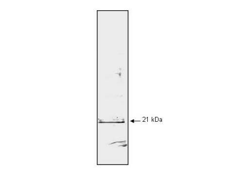
Mab anti-Human p21WAF1 antibody (clone WA-1) is shown to detect human p21 by western blot. Detection occurs after 10 μg of a HeLa whole cell lysate (p/n W09-000-364) is loaded per lane. The blot was incubated with a 1:1,000 dilution of Mab anti-Human p21WAF1 at room temperature for 30 min followed by detection using IRDye?800 labeled Goat-a-Mouse IgG [H&L] (p/n 610-132-121) diluted 1:5,000. A single band corresponding to human p21WAF1 is detected at ~21 kDa when compared with known molecular weight standards (not shown). The antibody may be used to detect endogenous human p21WAF1. IRDye?800 fluorescence image was captured using the OdysseyR Infrared Imaging System developed by LI-COR. IRDye is a trademark of LI-COR, Inc. Other detection systems will yield similar results.
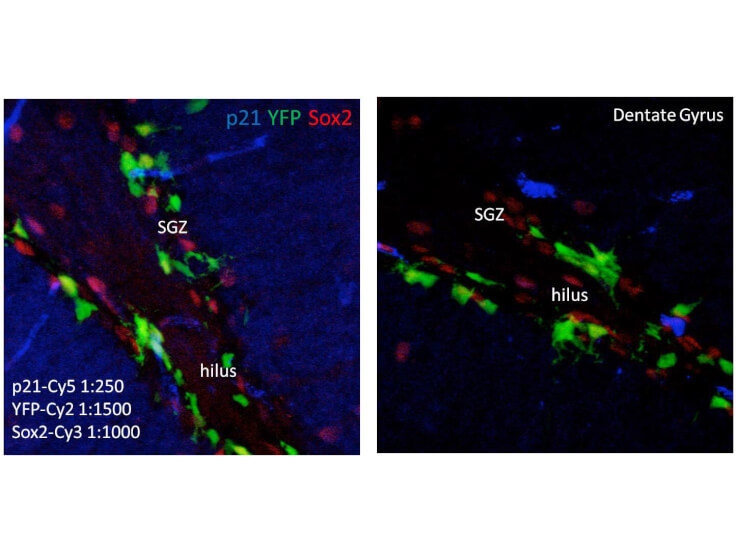
Immunofluorescence of Mouse anti-p21 antibody. Tissue: human brain. Fixation: free-floating. Antigen retrieval: not required. Primary antibody: anti-human-p21 antibody at 1:250 for 1 h at RT. Co-stained with YFP and Sox2 antibodies. Localization: p21 is nuclear and cytoplasmic. Staining: p21 as precipitated blue with Cy5, YFP as precipitated green with Cy2, and Sox2 as precipitated red with Cy3. z‐stacks from confocal expressed as one composite focal plain.
|
|
|
|
Mab anti-Human p21WAF1 antibody (clone WA-1) is shown to detect human p21 by western blot. Detection occurs after 10 μg of a HeLa whole cell lysate (p/n W09-000-364) is loaded per lane. The blot was incubated with a 1:1,000 dilution of Mab anti-Human p21WAF1 at room temperature for 30 min followed by detection using IRDye?800 labeled Goat-a-Mouse IgG [H&L] (p/n 610-132-121) diluted 1:5,000. A single band corresponding to human p21WAF1 is detected at ~21 kDa when compared with known molecular weight standards (not shown). The antibody may be used to detect endogenous human p21WAF1. IRDye?800 fluorescence image was captured using the OdysseyR Infrared Imaging System developed by LI-COR. IRDye is a trademark of LI-COR, Inc. Other detection systems will yield similar results.
|
|
| 別品名 |
mouse anti-p21/WAF1 antibody, Cyclin-dependent kinase inhibitor 1A (p21, CIP1) antibody, DNA Synthesis Inhibitor antibody, MDA 6 antibody, MDA6 antibody, Melanoma Differentiation Associated Protein 6 antibody
|
| 交差種 |
Human
|
| 適用 |
Western Blot
Enzyme Linked Immunosorbent Assay
Immuno Fluorescence
|
| 免疫動物 |
Mouse
|
| クローン |
WA-1
|
| 抗体クラス |
IgG1κ
|
| 標識物 |
Unlabeled
|
| 精製度 |
Ig fraction - Protein A
|
| GENE ID |
1026
|
| Accession No.(Gene/Protein) |
NP_510867.1, P38936
|
| Gene Symbol |
CDKN1A
|
| 参考文献 |
Yeh, C. H. and Shatkin, A.J. (1995). A cis-acting element in Rous sarcoma virus long terminal repeat required for promoter repression by Hela nuclear protein p21. J. Biol. Chem. 270:15815-15820 Joshi, U. S. et al. (1998). Inhibition of tumor cell growth by p21WAF1 adenoviral gene transfer in lung cancer. Cancer Gene Ther. 5: 183-191. Yang, Z.Y. et al. (1995) The p21 cyclin-dependent kinase inhibitor suppresses tumorigenicity in vivo. Nat Med 1(10):1052-1056 Kagawa, S. et al. (1997) p53 expression overcomes p21WAF1/CIP1-mediated G1 arrest and induces apoptosis in human cancer cells. Oncogene 15(16):1903-1909. Waldman, et al. (1995) p21 is necessary for the p53-mediated G1 arrest in human cancer cells. Cancer Res 55(22):5187-5190. Kovalic, J. et al., (1996) Int.J.Oncol. 9, suppl., 835. Stephane Supiot, Richard P. Hill and Robert G. Bristow (2008) Nutlin-3 radiosensitizes hypoxic prostate cancer cells independent of p53. Mol Cancer Ther 7; 993.
|
| [注意事項] |
濃度はロットによって異なる可能性があります。メーカーDS及びCoAからご確認ください。
|
|
| メーカー |
品番 |
包装 |
|
RKL
|
200-301-171
|
100 UG
|
※表示価格について
| 当社在庫 |
なし
|
| 納期目安 |
約10日
|
| 保存温度 |
-20℃
|
|
※当社では商品情報の適切な管理に努めておりますが、表示される法規制情報は最新でない可能性があります。
また法規制情報の表示が無いものは、必ずしも法規制に非該当であることを示すものではありません。
商品のお届け前に最新の製品法規制情報をお求めの際はこちらへお問い合わせください。
|
※当社取り扱いの試薬・機器製品および受託サービス・創薬支援サービス(納品物、解析データ等)は、研究用としてのみ販売しております。
人や動物の医療用・臨床診断用・食品用としては、使用しないように、十分ご注意ください。
法規制欄に体外診断用医薬品と記載のものは除きます。
|
|
※リンク先での文献等のダウンロードに際しましては、掲載元の規約遵守をお願いします。
|
|
※CAS Registry Numbers have not been verified by CAS and may be inaccurate.
|
