|
※サムネイル画像をクリックすると拡大画像が表示されます。
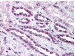
Rockland's anti-p65 pS276 antibody was diluted 1:500 to detect p65 in human kidney tissue. Tissue was formalin fixed and paraffin embedded. No pre-treatment of sample was required. The image shows the localization of antibody as the precipitated red signal, with a hematoxylin purple nuclear counter stain.
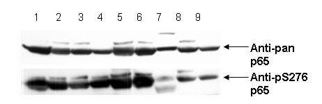
Anti-pS276 shows phospho p65 staining in carcinoma cells. Western blot of total protein lysates from various human head and neck tumors shows phospho p65 staining in tumor cell lines using phospho specific polyclonal anti-human pS276 p65. Lanes 1-6 contain protein lysates from human squamal carcinoma cell lines. Lane 7 is a protein lysate from a primary culture of human keratinocytes and does not show significant levels of phosphorylated p65. Lane 8 contains protein lysate from ATCC SCC9 cells (also a head and neck squamal carcinoma). Lane 9 contains lysate from EGF-induced human derived A431 cells. Lane 10 (not shown) contains a molecular weight standard. Concurrent staining with anti-beta microtubulin (not shown) was used to confirm equal protein loading in all lanes. HRP conjugated Gt-anti-Rabbit IgG was used to develop the blot using a chemiluminescent detection method. Other detection methods will yield similar results. Data contributed by Yu, M., NIH, personal communication.
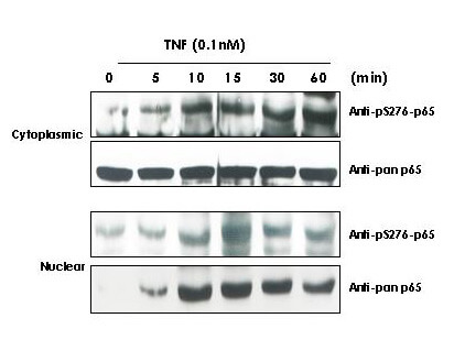
TNF Induces phosphorylation of p65 in KBM-5 cells. Cytoplasmic and nuclear protein lysates prepared after 0, 5, 10, 15, 30 and 60 minutes of 0.1 nM TNF treatment of KBM-5 cells shows inducible phosphorylation using phospho specific polyclonal anti-human pS276 p65. Rockland Immunochemical's pan reactive anti p65 (code# 100-4165) was used a control to show the presence of total p65 in both the cytoplasmic and nuclear extracts. Phosphorylation of p65 occurs after approximately 10 min of TNF exposure. Migration of phosphorylated p65 into the nucleus occurs within a similar time frame. HRP conjugated Gt-anti-Rabbit IgG was used to develop the blot using a chemi -luminescent detection method. Other detection methods will yield similar results. Personal Communication, Aggarwal BB
|
|
|
|
Rockland's anti-p65 pS276 antibody was diluted 1:500 to detect p65 in human kidney tissue. Tissue was formalin fixed and paraffin embedded. No pre-treatment of sample was required. The image shows the localization of antibody as the precipitated red signal, with a hematoxylin purple nuclear counter stain.
|
|
| 別品名 |
rabbit anti-NFkB p65 pS276 Antibody, rabbit anti-RelA pS276 Antibody, NFKB, nfkb, NF-kB, NF-kappaB, NFkappaB, Anti-NF-kB antibody
|
| 交差種 |
Human
|
| 適用 |
Western Blot
Enzyme Linked Immunosorbent Assay
Immunohistochemistry
|
| 免疫動物 |
Rabbit
|
| 抗原部位 |
QLRRPpSDRELSC
|
| 標識物 |
Unlabeled
|
| 精製度 |
Serum
|
| 翻訳後修飾 |
リン酸化
|
| GENE ID |
5970
|
| Accession No.(Gene/Protein) |
223468676, Q04206
|
| Gene Symbol |
RELA
|
| 参考文献 |
[Pub Med ID]27471270
|
| [注意事項] |
濃度はロットによって異なる可能性があります。メーカーDS及びCoAからご確認ください。
|
|
| メーカー |
品番 |
包装 |
|
RKL
|
100-401-264
|
100 UL
|
※表示価格について
| 当社在庫 |
なし
|
| 納期目安 |
約10日
|
| 保存温度 |
-20℃
|
|
※当社では商品情報の適切な管理に努めておりますが、表示される法規制情報は最新でない可能性があります。
また法規制情報の表示が無いものは、必ずしも法規制に非該当であることを示すものではありません。
商品のお届け前に最新の製品法規制情報をお求めの際はこちらへお問い合わせください。
|
※当社取り扱いの試薬・機器製品および受託サービス・創薬支援サービス(納品物、解析データ等)は、研究用としてのみ販売しております。
人や動物の医療用・臨床診断用・食品用としては、使用しないように、十分ご注意ください。
法規制欄に体外診断用医薬品と記載のものは除きます。
|
|
※リンク先での文献等のダウンロードに際しましては、掲載元の規約遵守をお願いします。
|
|
※CAS Registry Numbers have not been verified by CAS and may be inaccurate.
|
