|
※サムネイル画像をクリックすると拡大画像が表示されます。
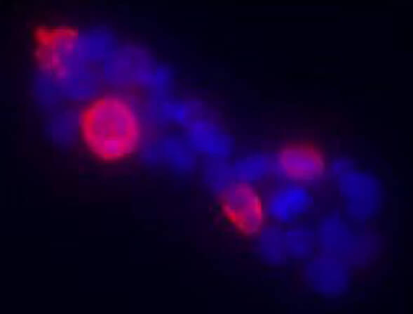
Immunofluorescence of Anti-Gli-1 antibody. HEK293T cells were transiently transfected with Gli-1 (murine). Primary Antibody: Rockland’s Anti-Gli-1 antiserum (rabbit) was added 1:400. Secondary Antibody: fluorescent labeled anti-rabbit IgG. Personal communication, Tom Curran, Children’s Hospital of Philadelphia, Philadelphia, PA. Detection of mouse Gli-1 present in transfected 293T cells (red).
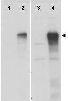
Western blot using Rockland's anti-Gli-1 antibody shows detection of a band at ~150 kDa corresponding to Gli-1 present in HEK293T whole cell lysate transiently transfected with Gli-1 (lane 2). Mock 293T cell lysates and 293T cell lysates transfected with Gli-2 and Gli-3 show no staining (lanes 1, 3 and 4 respectively). Lysates were separated by SDS-PAGE and transferred to nitrocellulose. After blocking, the membrane was probed with Anti-Gli-1 antiserum diluted 1:5,000, followed by HRP conjugated donkey anti-rabbit (Rockland p/n 611-7302). Personal communication, Tom Curran, Children’s Hospital of Philadelphia, Philadelphia, PA.
Western blot using Rockland's anti-Gli-1 antibody shows detection of a band at ~150 kDa (arrowhead) corresponding to human Gli-1 present in transfected 293T cell lysates (lanes 2 and 4). Mock 293T cell lysates with vector only show no staining (lanes 1 and 3). Lysates were separated by SDS-PAGE and transferred to nitrocellulose. After blocking the membrane was probed with the primary antibody diluted to 1:8,000 (lanes 1 and 2) or 1:4,000 (lanes 3 and 4). Molecular weight estimation was made by comparison to MW markers. Personal communication, Hiro Kimura, St. Jude Children's Research Hospital, Memphis, TN.
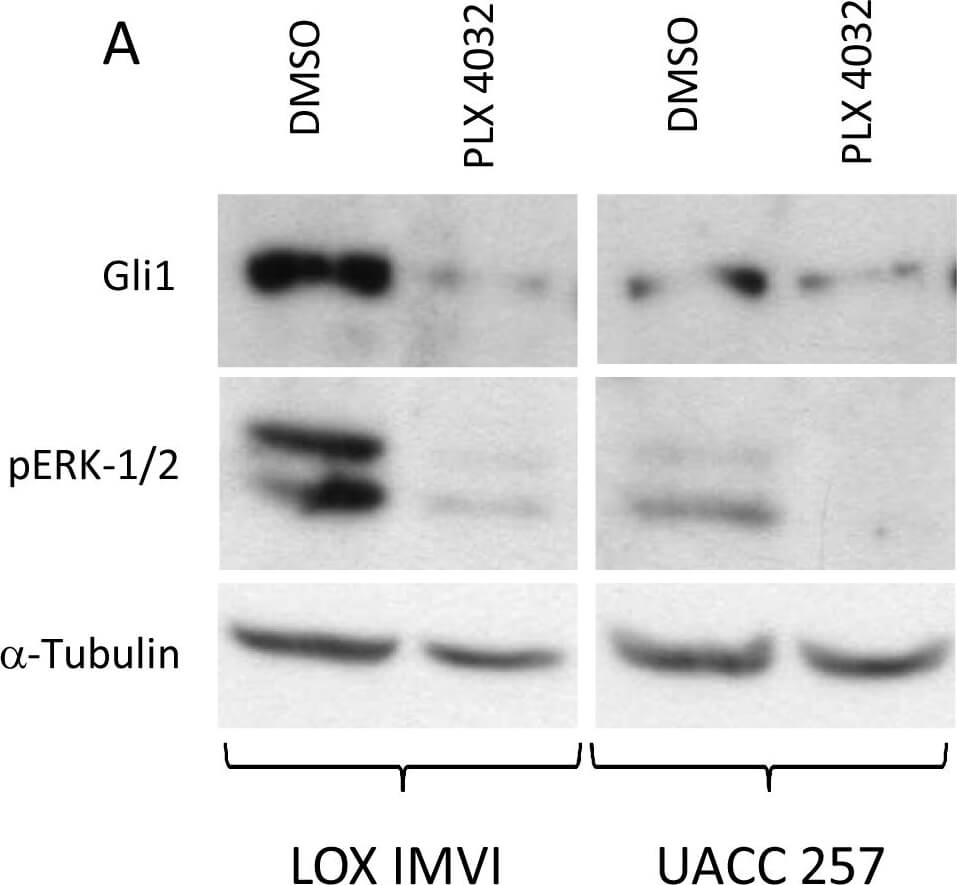
Inhibition of BRAFV600E, expression of GLI1 and SHH-GLI pathway inhibition by NVP-LDE225 in human melanoma cells in vitro. A) LOX IMVI and UACC 257 with PLX-4032 at the dose of 1 μM for 24 hr. subsequently protein lysates prepared and subjected to WB analysis for the expression of GLI1 and phospho-ERK1/2. B) Effect of NVP-LDE225 on PTCH1 promoter. In total, 1 μg of PTCH1 pGL3b-hPTCH1-prom-wt or pGL3b-hPTCH1-prom-mut luciferase construct and reporter were cotransfected into LOX IMVI cells. Cells were subsequently treated with 10 μM of NVP-LDE225 or cyclopamine for 4 hours (time point selected base on kinetic experiments). Fold activation was calculated relative to cells transfected with 3 μg of pB-actin-RL. One representative experiment of 2 is shown. Figure provided by CiteAb. Source: PLoS One, PMID: 23935925.
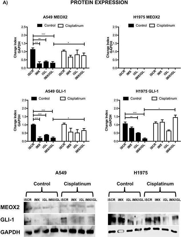
Inducible MEOX2-GLI1 axis expression was involved in cellular migration and cellular proliferation in lung cancer cells(A) A549 and H1975 lung cancer cells demonstrated an inducible GLI-1 protein expression pattern following treatment with 8 μM cisplatinum and reduced GLI-1 inducible expression following the application of specific anti-MEOX2 siRNA and/or anti-MEOX2 siRNA plus anti-GLI1 siRNA in the presence of 8 μM cisplatinum. Western blot statistical analyses, assessed via quantitative densitometry, were performed to determine *p?0.05 by one-way ANOVA and Dunnett's test for multiple comparisons to identify significant differences with respect to controls. Student's t-test was performed to identify significant differences between control and cisplatinum treatment. Quantification analyses were normalized to scrambled siRNA as a negative control for gene silencing. Images were obtained using a C-DIGIT device (LICOR), and pixel quantification and data analyses were carried out using Image Studio software. Total pixel intensity for each specific protein product was normalized to GAPDH. (B) Cell culture images and graphs showing the quantitative analysis of cellular migration as a percentage (transwell migration assays) indicated significant MEOX2 and GLI-1 protein-dependent functions following the individual and combined application of anti-MEOX2 and anti-GLI1 siRNAs in A549, NH2347 and H1975 lung adenocarcinoma cells; **p?0.005 and ***p?0.0001 based on one-way ANOVA and Dunnett's multiple comparisons test. (C) Cell culture images and graphs showing the quantitative analysis of cellular proliferation (clonogenic assays) indicated significant MEOX2 and GLI-1 protein-dependent functions following the individual and combined application of anti-MEOX2 and anti-GLI1 siRNAs in A549, NH2347 and H1975 lung adenocarcinoma cells; **p?0.005 and ***p?0.0001 based on one-way ANOVA and Dunnett's multiple comparisons test. Transwell migration index and colony growth (clonogenic as
|
|
|
|
Immunofluorescence of Anti-Gli-1 antibody. HEK293T cells were transiently transfected with Gli-1 (murine). Primary Antibody: Rockland’s Anti-Gli-1 antiserum (rabbit) was added 1:400. Secondary Antibody: fluorescent labeled anti-rabbit IgG. Personal communication, Tom Curran, Children’s Hospital of Philadelphia, Philadelphia, PA. Detection of mouse Gli-1 present in transfected 293T cells (red).
|
|
| 別品名 |
rabbit anti-Gli-1 Antibody, rabbit anti-Gli1 Antibody, Zinc finger protein GLI1 antibody, Glioma-associated oncogene antibody, Oncogene GLI antibody
|
| 交差種 |
Human
Mouse
|
| 適用 |
Western Blot
Enzyme Linked Immunosorbent Assay
Immuno Fluorescence
|
| 免疫動物 |
Rabbit
|
| 抗原部位 |
a.a.805-820
|
| 標識物 |
Unlabeled
|
| 精製度 |
Serum
|
| GENE ID |
14632
|
| Accession No.(Gene/Protein) |
4885279, P47806
|
| Gene Symbol |
Gli1
|
| 参考文献 |
[Pub Med ID]21995814
|
| [注意事項] |
濃度はロットによって異なる可能性があります。メーカーDS及びCoAからご確認ください。
|
|
| メーカー |
品番 |
包装 |
|
RKL
|
100-401-223
|
100 UL
|
※表示価格について
| 当社在庫 |
なし
|
| 納期目安 |
約10日
|
| 保存温度 |
-20℃
|
|
※当社では商品情報の適切な管理に努めておりますが、表示される法規制情報は最新でない可能性があります。
また法規制情報の表示が無いものは、必ずしも法規制に非該当であることを示すものではありません。
商品のお届け前に最新の製品法規制情報をお求めの際はこちらへお問い合わせください。
|
※当社取り扱いの試薬・機器製品および受託サービス・創薬支援サービス(納品物、解析データ等)は、研究用としてのみ販売しております。
人や動物の医療用・臨床診断用・食品用としては、使用しないように、十分ご注意ください。
法規制欄に体外診断用医薬品と記載のものは除きます。
|
|
※リンク先での文献等のダウンロードに際しましては、掲載元の規約遵守をお願いします。
|
|
※CAS Registry Numbers have not been verified by CAS and may be inaccurate.
|
