
| 別品名 |
G2/mitotic-specific cyclin-B1
|
| 種由来 |
Human
|
| 標識物 |
Unlabeled
|
| 精製度 |
Serum
|
| 適用 |
Western Blot
Immunohistochemistry
|
| 免疫動物 |
Rabbit
|
| 交差種 |
Human
|
| GENE ID |
891
|
| Accession No.(Gene/Protein) |
P14635
|
| Gene Symbol |
CCNB1
|
| 形状 |
滅菌済み液状品
|
| 参考文献 |
[Pub Med ID]24123312
|
| [注意事項] |
濃度はロットによって異なる可能性があります。メーカーDS及びCoAからご確認ください。
|
|
※サムネイル画像をクリックすると拡大画像が表示されます。
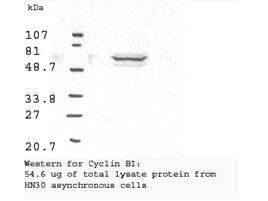
Western blot analysis using Rockland's Anti Cyclin B1 antibody shows detection of human Cyclin B1 present in asynchronous HN30 cell lysates.? HN30 cells are from head and neck cancer tumors that over express cyclin B1 and D1. Comparison to a molecular weight marker indicates a band of ~62 kDa corresponding to the expected molecular weight for the protein.? The blot was incubated with a 1:500 dilution of the antibody for 1 h at room temperature.? Detection occurred using a 1:10,000 of HRP conjugated Goat a Rabbit IgG 611 103 122 and chemiluminescence reagent with a 1 min exposure time.? Other detection systems will yield similar results. Personal communication, Luca Cote, Temple University, Philadelphia, PA.
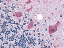
Rockland's anti-Cyclin B1 antibody was diluted 1:500 to detect Cyclin B1 in human brain cerebellum tissue. Tissue was formalin fixed and paraffin embedded. No pre-treatment of sample was required. The image shows the localization of antibody as the precipitated red signal, with a hematoxylin purple nuclear counter stain.
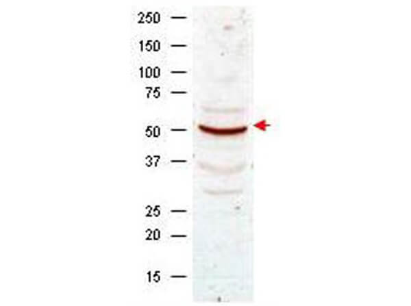
Western blot analysis using Rockland's anti-Cyclin B1 antibody shows detection of Cyclin B1 present in asynchronous HeLa cell lysates. Comparison to a molecular weight marker indicates a band of ~55 kDa corresponding to human Cyclin B1 (arrowhead). Approximately 50 ug of lysate was loaded on to a 7% SDS-PAGE gel for separation. After transfer to nitrocellulose, the blot was incubated with a 1:500 dilution of the antibody for 1 h at room temperature. Detection occurred using a 1:10,000 of HRP conjugated Goat-a-Rabbit IgG (p/n 611-103-122). Personal communication, Luca D'Agostino, Temple University, Philadelphia, PA.
|
|
|
|
Western blot analysis using Rockland's Anti Cyclin B1 antibody shows detection of human Cyclin B1 present in asynchronous HN30 cell lysates.? HN30 cells are from head and neck cancer tumors that over express cyclin B1 and D1. Comparison to a molecular weight marker indicates a band of ~62 kDa corresponding to the expected molecular weight for the protein.? The blot was incubated with a 1:500 dilution of the antibody for 1 h at room temperature.? Detection occurred using a 1:10,000 of HRP conjugated Goat a Rabbit IgG 611 103 122 and chemiluminescence reagent with a 1 min exposure time.? Other detection systems will yield similar results. Personal communication, Luca Cote, Temple University, Philadelphia, PA.
|
|
|
| メーカー |
品番 |
包装 |
|
RKL
|
100-401-152
|
100 UL
|
※表示価格について
| 当社在庫 |
なし
|
| 納期目安 |
約10日
|
| 保存温度 |
-20℃
|
|

