|
※サムネイル画像をクリックすると拡大画像が表示されます。
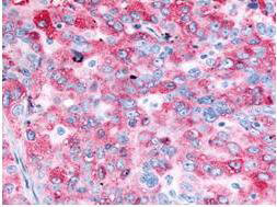
Immunohistochemistry of Anti-EGFR Antibody with positive staining.
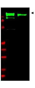
Western blot using Rockland's anti-EGFR antibody. Lane 1: unstimulated A431 whole cell lysates (p/n W09-000-361). Lane 2: EGF stimulated A431 whole cell lysates (p/n W09-000-362). Shows detection of a band at ~170 kDa corresponding to human EGFR present in unstimulated and stimulated (50 ng/ml for 15 min) lysates (arrowhead). Loaded: 30μg lysate was resolved on a 4-20% Tris-Glycine gel by SDS-PAGE and transferred onto nitrocellulose. Primary Antibody: Anti-EGFR at 1:1,000 overnight at 4° C. Secondary Antibody: IRDyeR 800 conjugated Gt-a-Rabbit IgG (H&L) MX10 (p/n 611-132-122) at 1:10,000 dilution of for 45 min at room temperature (800 nm channel, green). Molecular weight estimation was made by comparison to prestained MW markers in lane M (700 nm channel, red). IRDyeR 800 fluorescence image was captured using the OdysseyR Infrared Imaging System developed by LI-COR. IRDye is a trademark of LI-COR, Inc. Other detection systems will yield similar results.
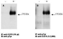
Combined immunoprecipitation and western blot using anti-EGFR antibody. Lysates were prepared from GN4 rat liver epithelial cells both with (+) EGF treatment for 15' at 100 ng/ml and without (-) the addition of EGF. The combination of immunoprecipitation and western blotting was performed using the anti-EGFR antibody for immunoprecipitation (10 μL) followed by western blot detection using an anti-phosphotyrosine antibody (Panel A). This was repeated in reverse order using a 1:2000 dilution of anti-EGFR for western blot (Panel B). Visualization occurred using an ECL system. Film exposure was approximately 1’. Other detection systems will yield similar results.
|
|
|
|
Immunohistochemistry of Anti-EGFR Antibody with positive staining.
|
|
| 別品名 |
rabbit anti-EGFR Antibody, rabbit anti-epidermal growth factor receptor antibody, Receptor tyrosine-protein kinase erbB-1 antibody, c-erbB-1 antibody
|
| 交差種 |
Human
Rat
|
| 適用 |
Western Blot
Immunohistochemistry
Immunoprecipitation
|
| 免疫動物 |
Rabbit
|
| 抗原部位 |
C-terminus
|
| 標識物 |
Unlabeled
|
| 精製度 |
Serum
|
| GENE ID |
1956
|
| Accession No.(Gene/Protein) |
29725609, P00533
|
| Gene Symbol |
EGFR
|
| 参考文献 |
[Pub Med ID]17130473
|
| [注意事項] |
濃度はロットによって異なる可能性があります。メーカーDS及びCoAからご確認ください。
|
|
| メーカー |
品番 |
包装 |
|
RKL
|
100-401-149
|
250 UL
|
※表示価格について
| 当社在庫 |
なし
|
| 納期目安 |
約10日
|
| 保存温度 |
-20℃
|
|
※当社では商品情報の適切な管理に努めておりますが、表示される法規制情報は最新でない可能性があります。
また法規制情報の表示が無いものは、必ずしも法規制に非該当であることを示すものではありません。
商品のお届け前に最新の製品法規制情報をお求めの際はこちらへお問い合わせください。
|
※当社取り扱いの試薬・機器製品および受託サービス・創薬支援サービス(納品物、解析データ等)は、研究用としてのみ販売しております。
人や動物の医療用・臨床診断用・食品用としては、使用しないように、十分ご注意ください。
法規制欄に体外診断用医薬品と記載のものは除きます。
|
|
※リンク先での文献等のダウンロードに際しましては、掲載元の規約遵守をお願いします。
|
|
※CAS Registry Numbers have not been verified by CAS and may be inaccurate.
|
