|
※サムネイル画像をクリックすると拡大画像が表示されます。
Figure 1 Western Blot Validation of CXCR4 in HeLa Cells
Loading: 15 μg of lysates per lane. Antibodies: 1009 (1 μg/mL), 1 h incubation at RT in 5% NFDM/TBST. Secondary: Goat anti-rabbit IgG HRP conjugate at 1:10000 dilution.
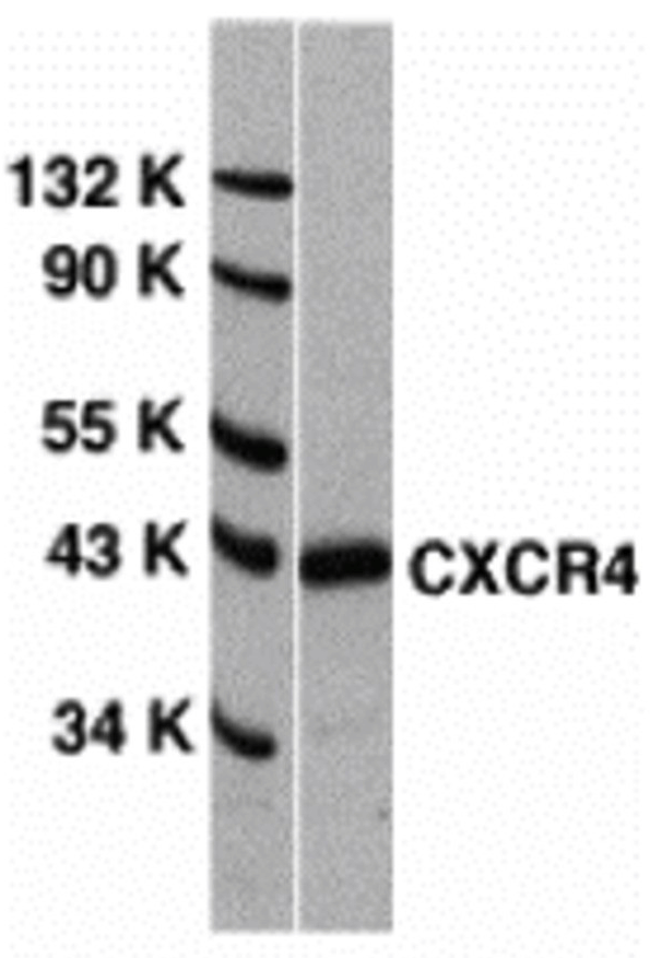
Figure 2 Independent Antibody Validation (IAV) via Protein Expression Profile in Cell Lines
Loading: 15 μg of lysates per lane. Antibodies: 1009 (1 μg/mL), 1012 (1 μg/mL), and beta-actin (1 μg/mL), 1 h incubation at RT in 5% NFDM/TBST. Secondary: Goat anti-rabbit IgG HRP conjugate at 1:10000 dilution.

Figure 3 Validation with CXCR4 siRNA Knockdown in HeLa Cells
HeLa cells were transfected with control siRNAs (lane 1) or CXCR4 siRNAs (lane 2) Loading: 10 μg of HeLa whole cell lysates per lane. Antibodies: 1009 (2 μg/mL), 1 h incubation at RT in 5% NFDM/TBST. Secondary: Goat anti-rabbit IgG HRP conjugate at 1:10000 dilution.

Figure 4 Animal Species Reactivity
Loading: Lysates/proteins at 20 μg per lane. Antibodies: 1009 (2 μg/mL) or 1012 (2 μg/mL). 1 h incubation at RT in 5% NFDM/TBST. Secondary: Goat anti-rabbit IgG HRP conjugate at 1:10000 dilution.
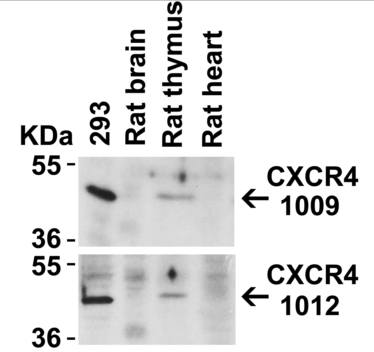
Figure 5 Recombinant Protein Test
Loading: CXCR4 partial recombinant protein (Novus Biologicals, Cat# H00007852-Q01). Lane 1: Anti-CXCR4 antibody (0.1 μg/mL) 1 h incubation at RT in 5% NFDM/TBST. Lane 2: Coomassie blue staining. Secondary: Goat anti-rabbit IgG HRP conjugate at 1:10000 dilution.
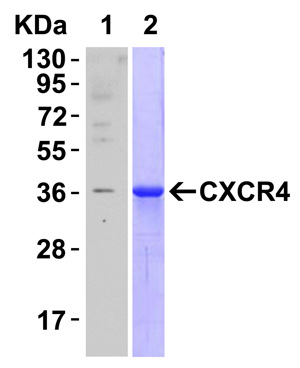
Figure 6 Immunofluorescence Validation of CXCR4 in HeLa Cells
Immunofluorescent analysis of 4% paraformaldehyde-fixed HeLa cells labeling CXCR4 with 1009 at 20 μg/mL, followed by goat anti-rabbit IgG secondary antibody at 1/500 dilution (red). Image showing both membrane and cytoplasmic staining on HeLa cells.
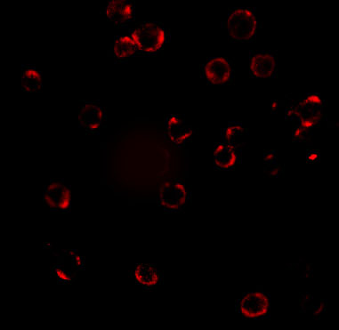
Figure 7 Flow Cytometry Validation of CXCR4 in HeLa Cells
Overlay histogram showing HeLa cells stained with 1009 (red line, 1μg/1x106 cells). 1 h incubation at 4℃ in 2% FBS/PBS. Followed by secondary antibody 488 goat anti-rabbit IgG (H+L) at 1/500 dilution for 1 h 4℃.
Isotype control antibody (Green line) was mouse IgG1 (1μg/1x106 cells) used under the same conditions.
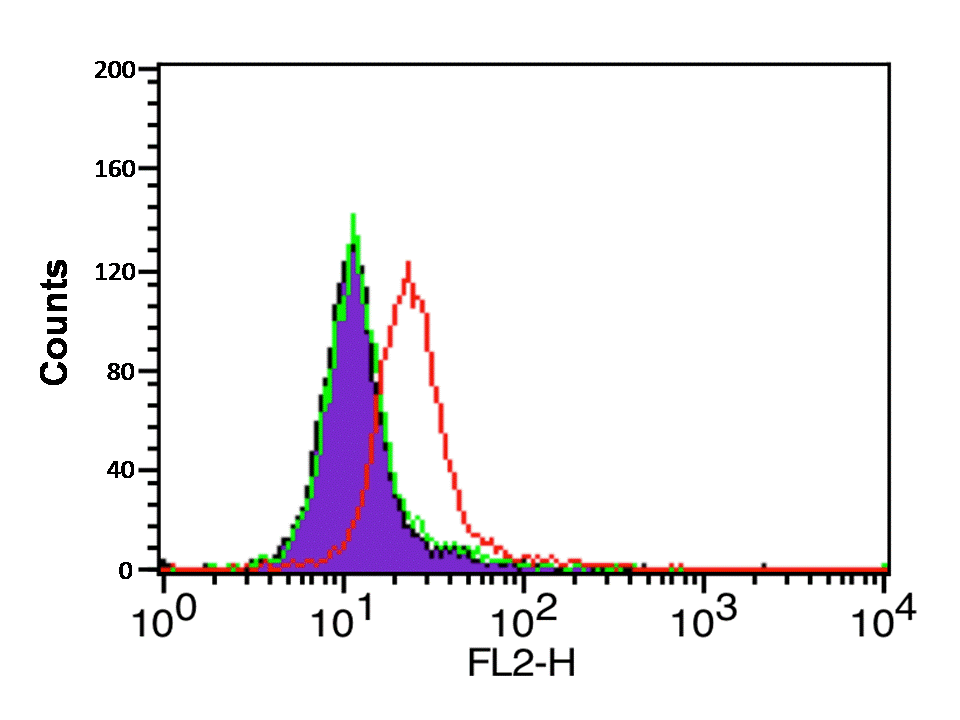
Figure 8 Immunohistochemistry Validation of CXCR4 in Human Spleen
Immunohistochemical analysis of paraffin-embedded human spleen tissue using anti-CXCR4 antibody (1009) at 5 μg/ml. Tissue was fixed with formaldehyde and blocked with 10% serum for 1 h at RT; antigen retrieval was by heat mediation with a citrate buffer (pH6). Samples were incubated with primary antibody overnight at 4℃. A Goat anti-rabbit IgG H&L (HRP) at 1/250 was used as secondary. Counter stained with Hematoxylin.
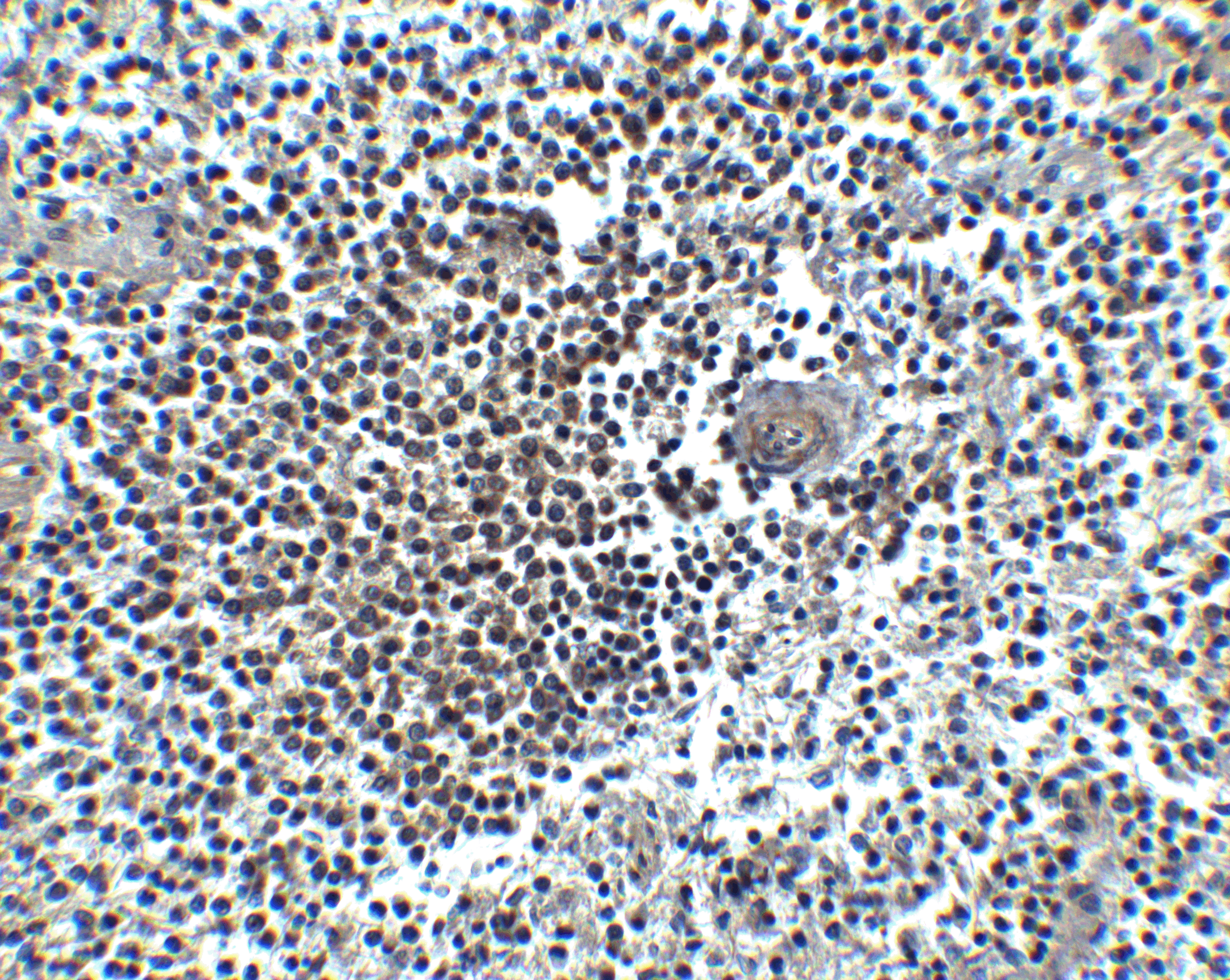
Figure 9 Immunocytochemistry Validation of CXCR4 in HeLa Cells
Immunocytochemical analysis of HeLa cells using anti-CXCR4 antibody (1009) at 2 μg/ml. Cells was fixed with formaldehyde and blocked with 10% serum for 1 h at RT; antigen retrieval was by heat mediation with a citrate buffer (pH6). Samples were incubated with primary antibody overnight at 4℃. A goat anti-rabbit IgG H&L (HRP) at 1/250 was used as secondary. Counter stained with Hematoxylin.
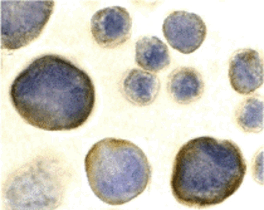
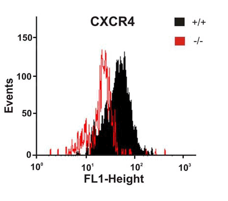
Figure 10 KO Validation of CXCR4 by Flow Cytometry (Odemis, et al., 2010)
Astrocytes from wild-type or CXCR4 knockout mice were stained with primary antibodies against CXCR4 and FITC-labeled secondary antibodies, and subsequently subjected to flow cytometry. CXCR4−/− astrocytes (red) showed loss of CXCR4 cell-surface expression compared with wild-type cells (black).
|

