|
※サムネイル画像をクリックすると拡大画像が表示されます。
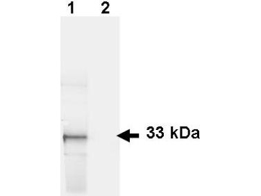
Anti-GFP Antibody - Western Blot. Western blot of GFP recombinant protein detected with polyclonal anti-GFP antibody. Lane 1 shows detection of a 33 kD band corresponding to a GFP containing recombinant protein (arrowhead) expressed in HeLa cells. Lane 2 shows no staining of a mock transfected HeLa cell lysate. A 4-12% Bis-Tris gradient gel was used for SDS-PAGE. The protein was transferred to nitrocellulose using standard methods. After blocking the membrane was probed with the primary antibody diluted to 1 ug/ml for 1 h at room temperature followed by washes and reaction with a 1:2500 dilution of IRDye 800 conjugated Donkey-a-Goat IgG [H&L] MX7 (. The IRDye 800 fluorescence image was captured using the Odyssey Infrared Imaging System developed by LI-COR. IRDye is a trademark of LI-COR, Inc. Other detection systems will yield similar results. This image was taken for the unconjugated form of this product. Other forms have not been tested.
Anti-GFP Antibody - Immunofluorescence Microscopy. polyclonal anti-GFP antibody at a 1:1000 dilution detects tau-GFP in cell bodies (large arrowhead) and axons of motorneurons (arrow) and interneurons (small arrowhead) in Drosophila melanogaster late stage embryonic central nervous system. Fluorochrome conjugated anti-Goat secondary antibody was used for detection at 1:300. Panel A shows a lateral view (ventral left) and Panels B and C show ventral views of whole mount embryos at 63x magnification (plus 2x digital zoom). In all panels, anterior is up. Personal Communication, Helmata Mistry, Washington University School of Medicine, St. Louis, MO. This image was taken for the unconjugated form of this product. Other forms have not been tested.

|
|
|
|
Anti-GFP Antibody - Western Blot. Western blot of GFP recombinant protein detected with polyclonal anti-GFP antibody. Lane 1 shows detection of a 33 kD band corresponding to a GFP containing recombinant protein (arrowhead) expressed in HeLa cells. Lane 2 shows no staining of a mock transfected HeLa cell lysate. A 4-12% Bis-Tris gradient gel was used for SDS-PAGE. The protein was transferred to nitrocellulose using standard methods. After blocking the membrane was probed with the primary antibody diluted to 1 ug/ml for 1 h at room temperature followed by washes and reaction with a 1:2500 dilution of IRDye 800 conjugated Donkey-a-Goat IgG [H&L] MX7 (. The IRDye 800 fluorescence image was captured using the Odyssey Infrared Imaging System developed by LI-COR. IRDye is a trademark of LI-COR, Inc. Other detection systems will yield similar results. This image was taken for the unconjugated form of this product. Other forms have not been tested.
|
|
| 別品名 |
GFP
|
| 種由来 |
Aequorea victoria
|
| 交差種 |
Aequorea victoria
|
| 免疫動物 |
Goat
|
| 抗体クラス |
IgG
|
| 標識物 |
Fluorescein Isothiocyanate
|
| 精製度 |
Affinity Purified
|
| Gene Symbol |
GFP
|
| その他 |
[適用]非標識抗体はWBとIFとFLISAに適用あり
|
|
| メーカー |
品番 |
包装 |
|
LSP
|
LS-C154186-1
|
1 MG
|
※表示価格について
| 当社在庫 |
なし
|
| 納期目安 |
約1ヶ月
|
| 保存温度 |
4℃
|
|
※当社では商品情報の適切な管理に努めておりますが、表示される法規制情報は最新でない可能性があります。
また法規制情報の表示が無いものは、必ずしも法規制に非該当であることを示すものではありません。
商品のお届け前に最新の製品法規制情報をお求めの際はこちらへお問い合わせください。
|
※当社取り扱いの試薬・機器製品および受託サービス・創薬支援サービス(納品物、解析データ等)は、研究用としてのみ販売しております。
人や動物の医療用・臨床診断用・食品用としては、使用しないように、十分ご注意ください。
法規制欄に体外診断用医薬品と記載のものは除きます。
|
|
※リンク先での文献等のダウンロードに際しましては、掲載元の規約遵守をお願いします。
|
|
※CAS Registry Numbers have not been verified by CAS and may be inaccurate.
|
