
| 別品名 |
Bcl-2 homologous antagonist/killer, Apoptosis regulator BAK, Bcl-2-like protein 7, Bcl2-L-7, BAK1, BAK, BCL2L7, CDN1
|
| 種由来 |
Human
|
| 標識物 |
Unlabeled
|
| 精製度 |
Affinity Purified
|
| 適用 |
Western Blot
Enzyme Linked Immunosorbent Assay
Immunohistochemistry
Flow Cytometry
Immunoprecipitation
|
| 免疫動物 |
Rabbit Mono
|
| 抗体クラス |
IgG
|
| クローン |
8D1
|
| 交差種 |
Human
|
| Accession No.(Gene/Protein) |
Q16611
|
| 形状 |
液状
|
|
※サムネイル画像をクリックすると拡大画像が表示されます。
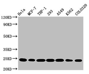
Western Blot
Positive WB detected in: Hela whole cell lysate, MCF-7 whole cell lysate, THP-1 whole cell lysate, 293 whole cell lysate, A549 whole cell lysate, K562 whole cell lysate, Colo320 whole cell lysate
All lanes: BAK1 antibody at 0.9ug/ml
Secondary
Goat polyclonal to rabbit IgG at 1/50000 dilution
Predicted band size: 24, 17 KDa
Observed band size: 24 KDa
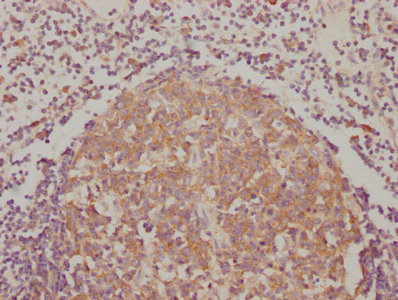
IHC image of CSB-RA624111A0HU diluted at 1:90 and staining in paraffin-embedded human lymph node tissue performed on a Leica BondTM system. After dewaxing and hydration, antigen retrieval was mediated by high pressure in a citrate buffer (pH 6.0). Section was blocked with 10% normal goat serum 30min at RT. Then primary antibody (1% BSA) was incubated at 4℃ overnight. The primary is detected by a biotinylated secondary antibody and visualized using an HRP conjugated SP system.
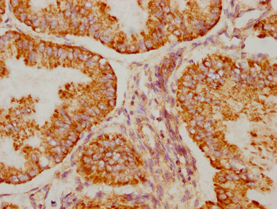
IHC image of CSB-RA624111A0HU diluted at 1:90 and staining in paraffin-embedded human endometrial cancer performed on a Leica BondTM system. After dewaxing and hydration, antigen retrieval was mediated by high pressure in a citrate buffer (pH 6.0). Section was blocked with 10% normal goat serum 30min at RT. Then primary antibody (1% BSA) was incubated at 4℃ overnight. The primary is detected by a biotinylated secondary antibody and visualized using an HRP conjugated SP system.
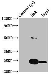
Immunoprecipitating BAK1 in HEK293 whole cell lysate
Lane 1: Rabbit control IgG instead of CSB-RA624111A0HU in HEK293 whole cell lysate.For western blotting, a HRP-conjugated Protein G antibody was used as the secondary antibody (1/2000)
Lane 2: CSB-RA624111A0HU (3ug) + HEK293 whole cell lysate (500ug)
Lane 3: HEK293 whole cell lysate (20ug)
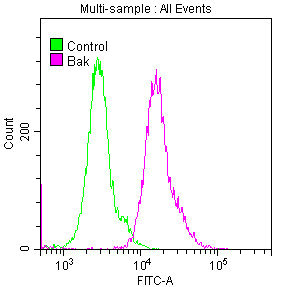
Overlay histogram showing Hela cells stained with CSB-RA624111A0HU (red line) at 1:50. The cells were fixed with 70% Ethylalcohol (18h) and then permeabilized with 0.3% Triton X-100 for 2 min. The cells were then incubated in 1x PBS /10% normal goat serum to block non-specific protein-protein interactions followed by primary antibody for 1 h at 4℃. The secondary antibody used was FITC goat anti-rabbit IgG (H+L) at 1/200 dilution for 1 h at 4℃. Control antibody (green line) was used under the same conditions. Acquisition of >10,000 events was performed.
|
|
|
|
Western Blot
Positive WB detected in: Hela whole cell lysate, MCF-7 whole cell lysate, THP-1 whole cell lysate, 293 whole cell lysate, A549 whole cell lysate, K562 whole cell lysate, Colo320 whole cell lysate
All lanes: BAK1 antibody at 0.9ug/ml
Secondary
Goat polyclonal to rabbit IgG at 1/50000 dilution
Predicted band size: 24, 17 KDa
Observed band size: 24 KDa
|
|
|
| メーカー |
品番 |
包装 |
|
CSB
|
CSB-RA624111A0HU
|
100 UL
|
※表示価格について
| 当社在庫 |
なし
|
| 納期目安 |
2週間程度
|
| 保存温度 |
-20℃
|
|

