
| 別品名 |
CCNY antibody; C10orf9 antibody; CBCP1 antibody; CFP1Cyclin-Y antibody; Cyc-Y antibody; Cyclin box protein 1 antibody; Cyclin fold protein 1 antibody; cyclin-X antibody
|
| 抗原部位 |
a.a.2-170
|
| 種由来 |
Human
|
| 標識物 |
Unlabeled
|
| 精製度 |
Ig fraction - Protein G
|
| 適用 |
Western Blot
Enzyme Linked Immunosorbent Assay
Immunohistochemistry
Immuno Fluorescence
|
| 免疫動物 |
Rabbit
|
| 抗体クラス |
IgG
|
| 交差種 |
Human
|
| Accession No.(Gene/Protein) |
Q8ND76
|
| 形状 |
液状
|
|
※サムネイル画像をクリックすると拡大画像が表示されます。
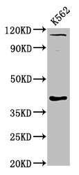
Western Blot
Positive WB detected in: K562 whole cell lysate
All lanes: CCNY antibody at 6μg/ml
Secondary
Goat polyclonal to rabbit IgG at 1/50000 dilution
Predicted band size: 40, 37, 34 kDa
Observed band size: 40 kDa
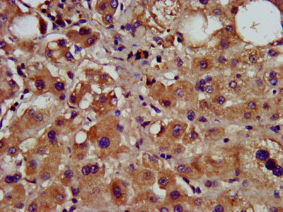
IHC image of CSB-PA847689LA01HU diluted at 1:600 and staining in paraffin-embedded human liver tissue performed on a Leica BondTM system. After dewaxing and hydration, antigen retrieval was mediated by high pressure in a citrate buffer (pH 6.0). Section was blocked with 10% normal goat serum 30min at RT. Then primary antibody (1% BSA) was incubated at 4°C overnight. The primary is detected by a biotinylated secondary antibody and visualized using an HRP conjugated SP system.
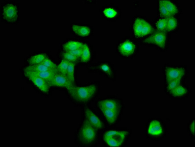
Immunofluorescence staining of HepG2 cells with CSB-PA847689LA01HU at 1:200, counter-stained with DAPI. The cells were fixed in 4% formaldehyde, permeabilized using 0.2% Triton X-100 and blocked in 10% normal Goat Serum. The cells were then incubated with the antibody overnight at 4°C. The secondary antibody was Alexa Fluor 488-congugated AffiniPure Goat Anti-Rabbit IgG(H+L).
|
|
|
|
Western Blot
Positive WB detected in: K562 whole cell lysate
All lanes: CCNY antibody at 6μg/ml
Secondary
Goat polyclonal to rabbit IgG at 1/50000 dilution
Predicted band size: 40, 37, 34 kDa
Observed band size: 40 kDa
|
|
|
| メーカー |
品番 |
包装 |
|
CSB
|
CSB-PA847689LA01HU
|
100 UG
|
※表示価格について
| 当社在庫 |
なし
|
| 納期目安 |
2週間程度
|
| 保存温度 |
-20℃
|
|

