|
※サムネイル画像をクリックすると拡大画像が表示されます。
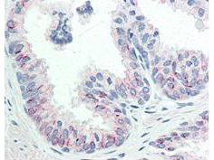
Immunohistochemistry of Mouse anti-AKT pS473 antibody. Tissue: human prostate tissue. Fixation: formalin fixed paraffin embedded. Antigen retrieval: not required. Primary antibody: AKT pS473 antibody at 20 ツオg/mL for 1 h at RT. Secondary antibody: Dako's Techmate streptavidin-biotin reagents at 1:10,000 for 45 min at RT. Localization: AKT pS473 is nuclear and occasionally cytoplasmic. Staining: AKT pS473 as precipitated red signal with hematoxylin purple nuclear counterstain.
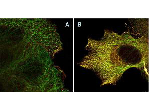
Immunofluorescence Microscopy of Mouse Anti-AKTpS473 antibody using STED nanoscopy to evaluate AKT activation and migration. Tissue: A431 cells. Antigen retrieval: Panel A: serum starved,unstimulated cells. Panel B: serum starved, EGF stimulated for 15 mins. A massive increase in AKT-pS473 activation, as measured by intensity signal, peaked at 15 minutes and was associated with depolymerized tubulin. Staining: Panel A shows STED data (AKT-pS473, red channel) collected simultaneously with confocal signal (a-tubulin, green channel). Upon stimulation of cells with EGF, a rapid activation of AKT is observed (Panel B) along with a coincident change in the tubulin organization (yellow signal), as well as an extensive cell shape-change (cell membrane folding) and accumulation of AKTpS473 at the cell periphery.
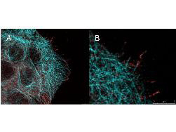
High resolution STED immunofluorescence nanoscopy of Mouse anti-AKT pS473 antibody. Tissue: A431 cells. The merge images (A) and at high magnification (B) show phosphorylated AKT colocalized with the distal microtubules. Fixation: 4% paraformaldehyde for 5 min and after washes blocked with 10% NGS/0.2% Triton X-100 for 30 min. Antigen retrieval: serum deprivation for 12 h. Primary antibody: AKT pS473 antibody at 10 ツオg/mL and a-tubulin (cyan) at 1.4 ツオg/mL for 1 h at RT. Secondary antibody: Atto 647N anti-Mouse IgG (ATTO TEC GmbH), and DyLight 488 anti-Rabbit IgG were used at 1.0 ツオg/mL for 1h at RT for indirect detection. Localization: AKT pS473 is in the cytoplasm and also organized at the periphery of the cell. Staining: AKT pS473 as red signal with bis-benzimide (blue) nuclear counterstain.
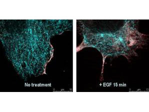
Immunofluorescence confocal microscopy of Mouse Anti-AKT pS473 antibody. Tissue: EGF treated A431 cells. Fixation: 0.5% PFA. Antigen retrieval: EGF 15 min. Primary antibody: AKT pS473 antibody at 10 ツオg/mL for 1 h at RT. Secondary antibody: DyLight 488 Goat anti-Rabbit IgG, MAb anti-AKT pS473, atto-647N anti-Mouse IgG (Active Motif). at 1:10,000 for 45 min at RT. Localization: AKT pS473 is nuclear and occasionally cytoplasmic. Staining: AKT pS473 as red signal with tubulin (cyan).
Western Blot of Mouse Anti-AKT pS473 antibody. Lane 1: non-phosphorylated AKT in untreated cells. Lane 2: phosphorylated AKT (indicated by arrowhead at ~56 kDa) on PDGF stimulated NIH/3T3 cell lysates. Load: 10 ツオg per lane. Primary antibody: AKT pS473 antibody at 1:10,000 in TBS with 0.05% Tween-20 with 1% BSA, for 1 h at 4ツー C. Secondary antibody: HRP conjugated Gt-a-Mouse IgG was used at a 1:20,000 dilution for 1 h at 4ツー C with FemtoMax enhanced chemiluminescent reagent Other band(s): none.

Western Blot of Mouse Anti-Akt pS473 antibody. Lane 1: unstimulated NIH/3T3 lysates contain inactive unphosphorylated Akt1, green band. Lane 2: PDGF stimulated NIH/3T3 lysate contains both inactive (green band) and activated phosphorylated Akt1 (red band). Load: 10 ツオg per lane. Primary antibody: rabbit anti-Akt (pan) and mouse anti-Akt pS473 specific antibodies at 1:400 for overnight at 4ツーC. Secondary antibody: DyLight 549 conjugated anti-rabbit IgG (green) and DyLight 649 conjugated anti-mouse IgG (red) secondary antibodies at 1:10,000 for 45 min at RT. Block: 5% BLOTTO overnight at 4ツーC.
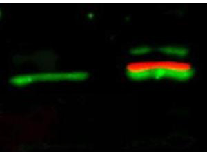
Western Blot of Mouse Anti-AKTpS473 antibody. Lane 1: A431 cells. Lane 2: A431 cells stimulated for 15 min with EGF. Load: 35 ツオg per lane. Primary antibody: AKTpS473 antibody at 1:400 for overnight at 4ツーC. Secondary antibody: DyLight649 Conjugated Anti-AKT pS473 Monoclonal Antibody p/n 200-343-268 at 1:10,000 for 45 min at RT. Block: Blocking Buffer for Fluorescent Western Blotting p/n MB-070 overnight at 4ツーC. Predicted/Observed size: 56kDa. Other band(s): none.

Western Blot of Mouse Anti-Akt pS473 antibody. A Lane 1) PDGF stimulated NIH 3T3 cells 10 ? Lane 2) NIH 3T3 cells 10 ? Lane 3) Hela whole cell lysate 10 ? (weak signal) B Lane 4) GST negative control protein 100 ng Lane 5) GST negative control protein 25 ng Lane 6) AKT 1 recombinant protein 100 ng Lane 7) AKT 1 recombinant protein 25 ng Block: 5% BSA overnight at 4?. Primary antibody: Monoclonal anti AKT antibody, used at 1:1000 for overnight at 4?. Secondary antibody: HRP Conjugated goat anti mouse lot 20121 1:25K for 45 min at RT. Detection : TMBM-100 for 20 minutes, rinsed with deionized water, dried and scanned on conventional flatbed scanner

|
|
|
|
Immunohistochemistry of Mouse anti-AKT pS473 antibody. Tissue: human prostate tissue. Fixation: formalin fixed paraffin embedded. Antigen retrieval: not required. Primary antibody: AKT pS473 antibody at 20 ツオg/mL for 1 h at RT. Secondary antibody: Dako's Techmate streptavidin-biotin reagents at 1:10,000 for 45 min at RT. Localization: AKT pS473 is nuclear and occasionally cytoplasmic. Staining: AKT pS473 as precipitated red signal with hematoxylin purple nuclear counterstain.
|
|
| 別品名 |
AKT1, AKT, AKT-1, C-AKT, Pan-AKT, PKB alpha, PKBalpha, Proto-oncogene c-Akt, Protein kinase B, Rac protein kinase alpha, PRKBA, Protein kinase B alpha, RAC-PK-alpha, PKB, PKB-ALPHA, RAC, RAC-ALPHA
|
| 種由来 |
Human
|
| 交差種 |
Human
Mouse
Rat
Monkey
|
| 適用 |
Western Blot
Enzyme Linked Immunosorbent Assay
Immunohistochemistry
Immuno Fluorescence
Flow Cytometry
Immunoprecipitation
|
| 免疫動物 |
Mouse
|
| クローン |
17F6.B11
|
| 抗体クラス |
IgG1κ
|
| 標識物 |
Unlabeled
|
| 精製度 |
Ig fraction - Protein A
|
| 翻訳後修飾 |
リン酸化
|
| GENE ID |
207
|
| Gene Symbol |
AKT1
|
|
| メーカー |
品番 |
包装 |
|
LSP
|
LS-C744816-25
|
25 UL
|
※表示価格について
| 当社在庫 |
なし
|
| 納期目安 |
約1ヶ月
|
| 保存温度 |
-20℃
|
|
※当社では商品情報の適切な管理に努めておりますが、表示される法規制情報は最新でない可能性があります。
また法規制情報の表示が無いものは、必ずしも法規制に非該当であることを示すものではありません。
商品のお届け前に最新の製品法規制情報をお求めの際はこちらへお問い合わせください。
|
※当社取り扱いの試薬・機器製品および受託サービス・創薬支援サービス(納品物、解析データ等)は、研究用としてのみ販売しております。
人や動物の医療用・臨床診断用・食品用としては、使用しないように、十分ご注意ください。
法規制欄に体外診断用医薬品と記載のものは除きます。
|
|
※リンク先での文献等のダウンロードに際しましては、掲載元の規約遵守をお願いします。
|
|
※CAS Registry Numbers have not been verified by CAS and may be inaccurate.
|


