|
※サムネイル画像をクリックすると拡大画像が表示されます。
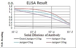
ELISA analysis of WTAP monoclonal antibody, clone 6B6B6.
Western blot analysis of Lane 1: HEK293 cell; Lane 2: WTAP-hIgGFc transfected HEK293 cell with WTAP monoclonal antibody.

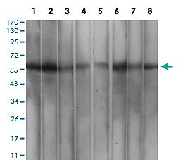
Western blot analysis of Lane 1: MCF-7 cell; Lane 2: Hela cell; Lane 3: K562 cell; Lane 4: Hek293 cell; Lane 5: A549 cell; Lane 6: HepG2 cell; Lane 7: Jurkat cell and Lane 8: Cos7 cell with WTAP monoclonal antibody.
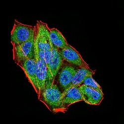
Immunocytochemical staining of Hela cells with WTAP monoclonal antibody (green). DRAQ5 fluorescent DNA dye (blue). Actin filaments labeled with Alexa Fluor-555 phalloidin (red).
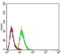
Flow cytometric analysis of A549 cells with WTAP monoclonal antibody (green) and negative control (red).
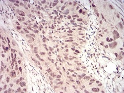
Immunohistochemical staining of paraffin-embedded cervical cancer tissues with WTAP monoclonal antibody.
|

