
| 抗原部位 |
a.a.192-212
|
| 種由来 |
Bovine
|
| 精製度 |
Ig fraction - Protein G
|
| 適用 |
Western Blot
Immuno Fluorescence
Immunoprecipitation
|
| 免疫動物 |
Mouse
|
| クローン |
KR-10
|
| 交差種 |
Human
Mouse
Rat
|
| Gene Symbol |
Kdelr1
|
| 形状 |
液状
|
| その他 |
[Uniprot ID]Q99JH8
|
|
※サムネイル画像をクリックすると拡大画像が表示されます。
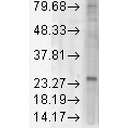
Figure 2. Western blot analysis of Kdelr1 using anti-Kdelr1 antibody (M07617-2).
Electrophoresis was performed on a 5-20% SDS-PAGE gel at 70V (Stacking gel) / 90V (Resolving gel) for 2-3 hours. The sample well of each lane was loaded with 50ug of sample under reducing conditions.
After Electrophoresis, proteins were transferred to a Nitrocellulose membrane at 150mA for 50-90 minutes. Blocked the membrane with 5% Non-fat Milk/ TBS for 1.5 hour at RT. The membrane was incubated with rabbit anti-Kdelr1 antigen affinity purified polyclonal antibody (Catalog # M07617-2) at 0.5 ug/mL overnight at 4°C, then washed with TBS-0.1%Tween 3 times with 5 minutes each and probed with a goat anti-Mouse IgG-HRP secondary antibody at a dilution of 1:10000 for 1.5 hour at RT. The signal is developed using an Enhanced Chemiluminescent detection (ECL) kit (Catalog # SA1021) with Tanon 5200 system. A specific band was detected for Kdelr1.
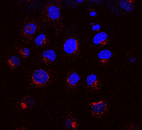
Immunocytochemistry/Immunofluorescence analysis using Mouse Anti-KDEL Receptor Monoclonal Antibody, Clone KR-10 (M07617-2) . Tissue: NRK cells. Species: Rat. Primary Antibody: Mouse Anti-KDEL Receptor Monoclonal Antibody (M07617-2) at 1:1000. Secondary Antibody: APC Goat Anti-Mouse (red) . Counterstain: DAPI (blue) nuclear stain. KR-10 staining red; DAPI staining blue. Merged images. Courtesy of: Institute of Mol. and Cell Bio, Singapore.
|
|
|
|
Figure 2. Western blot analysis of Kdelr1 using anti-Kdelr1 antibody (M07617-2).
Electrophoresis was performed on a 5-20% SDS-PAGE gel at 70V (Stacking gel) / 90V (Resolving gel) for 2-3 hours. The sample well of each lane was loaded with 50ug of sample under reducing conditions.
After Electrophoresis, proteins were transferred to a Nitrocellulose membrane at 150mA for 50-90 minutes. Blocked the membrane with 5% Non-fat Milk/ TBS for 1.5 hour at RT. The membrane was incubated with rabbit anti-Kdelr1 antigen affinity purified polyclonal antibody (Catalog # M07617-2) at 0.5 ug/mL overnight at 4°C, then washed with TBS-0.1%Tween 3 times with 5 minutes each and probed with a goat anti-Mouse IgG-HRP secondary antibody at a dilution of 1:10000 for 1.5 hour at RT. The signal is developed using an Enhanced Chemiluminescent detection (ECL) kit (Catalog # SA1021) with Tanon 5200 system. A specific band was detected for Kdelr1.
|
|
|
| メーカー |
品番 |
包装 |
|
BBT
|
M07617-2
|
100 UG
|
※表示価格について
|

