
| 抗原部位 |
a.a.750-850
|
| 種由来 |
Human
|
| 精製度 |
Ig fraction - Protein G
|
| 適用 |
Western Blot
Immunohistochemistry
|
| 免疫動物 |
Mouse
|
| クローン |
44C143
|
| 交差種 |
Human
Mouse
|
| Gene Symbol |
TLR8
|
| 形状 |
液状
|
| その他 |
[Uniprot ID]Q9NR97
|
|
※サムネイル画像をクリックすると拡大画像が表示されます。
Figure 2. IHC analysis of TLR8 using anti-TLR8 antibody (M01541).
TLR8 was detected in paraffin-embedded section. Heat mediated antigen retrieval was performed in citrate buffer (pH6, epitope retrieval solution) for 20 mins. The tissue section was blocked with 10% goat serum. The tissue section was then incubated with 1ug/ml rabbit anti-TLR8 Antibody (M01541) overnight at 4°C. Biotinylated goat anti Mouse IgG antibody was used as secondary antibody and incubated for 30 minutes at 37°C. The tissue section was developed using Strepavidin-Biotin-Complex (SABC)(Catalog # SA1021) with DAB as the chromogen.
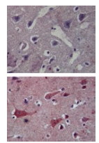
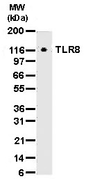
Figure 1. Western blot analysis of TLR8 using anti-TLR8 antibody (M01541).
Electrophoresis was performed on a 5-20% SDS-PAGE gel at 70V (Stacking gel) / 90V (Resolving gel) for 2-3 hours. The sample well of each lane was loaded with 50ug of sample under reducing conditions.
After Electrophoresis, proteins were transferred to a Nitrocellulose membrane at 150mA for 50-90 minutes. Blocked the membrane with 5% Non-fat Milk/ TBS for 1.5 hour at RT. The membrane was incubated with rabbit anti-TLR8 antigen affinity purified polyclonal antibody (Catalog # M01541) at 0.5 ug/mL overnight at 4°C, then washed with TBS-0.1%Tween 3 times with 5 minutes each and probed with a goat anti-Mouse IgG-HRP secondary antibody at a dilution of 1:10000 for 1.5 hour at RT. The signal is developed using an Enhanced Chemiluminescent detection (ECL) kit (Catalog # SA1021) with Tanon 5200 system. A specific band was detected for TLR8.
|
|
|
|
Figure 2. IHC analysis of TLR8 using anti-TLR8 antibody (M01541).
TLR8 was detected in paraffin-embedded section. Heat mediated antigen retrieval was performed in citrate buffer (pH6, epitope retrieval solution) for 20 mins. The tissue section was blocked with 10% goat serum. The tissue section was then incubated with 1ug/ml rabbit anti-TLR8 Antibody (M01541) overnight at 4°C. Biotinylated goat anti Mouse IgG antibody was used as secondary antibody and incubated for 30 minutes at 37°C. The tissue section was developed using Strepavidin-Biotin-Complex (SABC)(Catalog # SA1021) with DAB as the chromogen.
|
|
|
| メーカー |
品番 |
包装 |
|
BBT
|
M01541
|
100 UL
|
※表示価格について
| 当社在庫 |
なし
|
| 納期目安 |
1週間程度
|
| 保存温度 |
-20℃
|
|

