|
※サムネイル画像をクリックすると拡大画像が表示されます。
Immunofluorescence analysis of endoplasmic reticulum staining of mouse C2C12 myoblasts transfected with wild type mouse ADAM12 using KDEL (Grp78, Grp94) mAb (10C3).
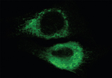
Figure 1. IHC analysis of Hspa5 using anti-Hspa5 antibody (M00955-2).
Hspa5 was detected in paraffin-embedded section. Heat mediated antigen retrieval was performed in citrate buffer (pH6, epitope retrieval solution) for 20 mins. The tissue section was blocked with 10% goat serum. The tissue section was then incubated with 1ug/ml rabbit anti-Hspa5 Antibody (M00955-2) overnight at 4°C. Biotinylated goat anti Mouse IgG antibody was used as secondary antibody and incubated for 30 minutes at 37°C. The tissue section was developed using Strepavidin-Biotin-Complex (SABC)(Catalog # SA1021) with DAB as the chromogen.

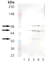
Figure 3. Western blot analysis of Hspa5 using anti-Hspa5 antibody (M00955-2).
Electrophoresis was performed on a 5-20% SDS-PAGE gel at 70V (Stacking gel) / 90V (Resolving gel) for 2-3 hours. The sample well of each lane was loaded with 50ug of sample under reducing conditions.
After Electrophoresis, proteins were transferred to a Nitrocellulose membrane at 150mA for 50-90 minutes. Blocked the membrane with 5% Non-fat Milk/ TBS for 1.5 hour at RT. The membrane was incubated with rabbit anti-Hspa5 antigen affinity purified polyclonal antibody (Catalog # M00955-2) at 0.5 ug/mL overnight at 4°C, then washed with TBS-0.1%Tween 3 times with 5 minutes each and probed with a goat anti-Mouse IgG-HRP secondary antibody at a dilution of 1:10000 for 1.5 hour at RT. The signal is developed using an Enhanced Chemiluminescent detection (ECL) kit (Catalog # SA1021) with Tanon 5200 system. A specific band was detected for Hspa5.
|

