|
※サムネイル画像をクリックすると拡大画像が表示されます。
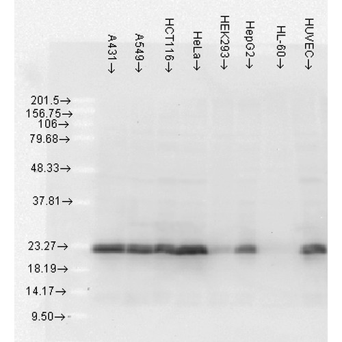
Figure 4. Western blot analysis of HSPB1 using anti-HSPB1 antibody (M00676-4).
Electrophoresis was performed on a 5-20% SDS-PAGE gel at 70V (Stacking gel) / 90V (Resolving gel) for 2-3 hours. The sample well of each lane was loaded with 50ug of sample under reducing conditions.
After Electrophoresis, proteins were transferred to a Nitrocellulose membrane at 150mA for 50-90 minutes. Blocked the membrane with 5% Non-fat Milk/ TBS for 1.5 hour at RT. The membrane was incubated with rabbit anti-HSPB1 antigen affinity purified polyclonal antibody (Catalog # M00676-4) at 0.5 ug/mL overnight at 4°C, then washed with TBS-0.1%Tween 3 times with 5 minutes each and probed with a goat anti-Mouse IgG-HRP secondary antibody at a dilution of 1:10000 for 1.5 hour at RT. The signal is developed using an Enhanced Chemiluminescent detection (ECL) kit (Catalog # SA1021) with Tanon 5200 system. A specific band was detected for HSPB1.
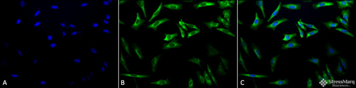
Immunocytochemistry/Immunofluorescence analysis using Mouse Anti-Hsp27 Monoclonal Antibody, Clone 5D12-A3 (M00676-4) . Tissue: Heat Shocked HeLa Cells. Species: Human. Fixation: 2% Formaldehyde for 20 min at RT. Primary Antibody: Mouse Anti-Hsp27 Monoclonal Antibody (M00676-4) at 1:100 for 12 hours at 4°C. Secondary Antibody: FITC Goat Anti-Mouse (green) at 1:200 for 2 hours at RT. Counterstain: DAPI (blue) nuclear stain at 1:40000 for 2 hours at RT. Localization: Cytoplasm. Nucleus. Magnification: 20x. (A) DAPI (blue) nuclear stain. (B) Anti-Hsp27 Antibody. (C) Composite. Heat Shocked at 42°C for 1h.
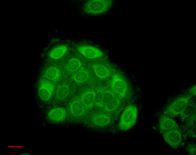
Immunocytochemistry/Immunofluorescence analysis using Mouse Anti-Hsp27 Monoclonal Antibody, Clone 5D12-A3 (M00676-4) . Tissue: HaCaT cells. Species: Human. Fixation: Cold 100% methanol for 10 minutes at -20°C. Primary Antibody: Mouse Anti-Hsp27 Monoclonal Antibody (M00676-4) at 1:100 for 1 hour at RT. Secondary Antibody: FITC Goat Anti-Mouse (green) at 1:50 for 1 hour at RT. Localization: Dull heterogeneous staining, some perinuclear, some nuclear and some cytoplasmic staining .
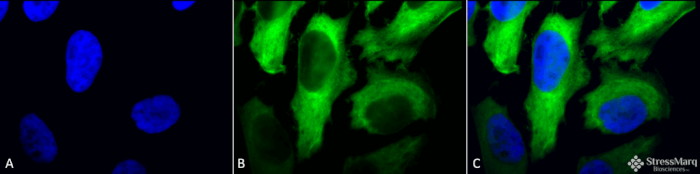
Immunocytochemistry/Immunofluorescence analysis using Mouse Anti-Hsp27 Monoclonal Antibody, Clone 5D12-A3 (M00676-4) . Tissue: Heat Shocked HeLa Cells. Species: Human. Fixation: 2% Formaldehyde for 20 min at RT. Primary Antibody: Mouse Anti-Hsp27 Monoclonal Antibody (M00676-4) at 1:100 for 12 hours at 4°C. Secondary Antibody: FITC Goat Anti-Mouse (green) at 1:200 for 2 hours at RT. Counterstain: DAPI (blue) nuclear stain at 1:40000 for 2 hours at RT. Localization: Cytoplasm. Nucleus. Magnification: 100x. (A) DAPI (blue) nuclear stain. (B) Anti-Hsp27 Antibody. (C) Composite. Heat Shocked at 42°C for 1h.
|

