
| 別品名 |
Histone H3.1;Histone H3/a;Histone H3/b;Histone H3/c;Histone H3/d;Histone H3/f;Histone H3/h;Histone H3/i;Histone H3/j;Histone H3/k;Histone H3/l;HIST1H3A;H3FA;HIST1H3B;H3FL;HIST1H3C;H3FC;HIST1H3D;H3FB;HIST1H3E;H3FD;HIST1H3F;H3FI;HIST1H3G;H3FH;HIST1H3H;H3FK;HIST1H3I;H3FF;HIST1H3J;H3FJ
|
| 種由来 |
Human
|
| 標識物 |
Unlabeled
|
| 精製度 |
Affinity Purified
|
| 適用 |
Western Blot
Immunohistochemistry
Immuno Fluorescence
Immunoprecipitation
Chromatin Immunoprecipitation
|
| 免疫動物 |
Rabbit Mono
|
| クローン |
AFF-8
|
| 交差種 |
Human
Mouse
Rat
|
| 翻訳後修飾 |
メチル化
|
| Accession No.(Gene/Protein) |
P68431
|
| Gene Symbol |
HIST1H3AHIST1H3BHIST1H3CHIST1H3DHIST1H3EHIST1H3FHIST1H3GHIST1H3HHIST1H3IHIST1H3J
|
| 分子量 |
15404 MW
|
| 形状 |
液状
|
|
※サムネイル画像をクリックすると拡大画像が表示されます。
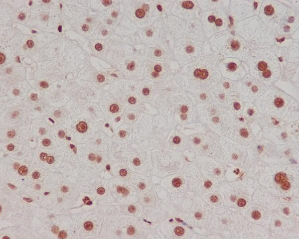
Immunohistochemical analysis of paraffin-embedded human liver, using Methyl-Histone H3 (di K4) Antibody(M12477-1)HIST1H3A was detected in paraffin-embedded tissue section. Heat mediated antigen retrieval was performed in citrate buffer (pH6, epitope retrieval solution) for 20 mins. The tissue section was blocked with 10% goat serum. The tissue section was then incubated with 1ug/ml rabbit anti-HIST1H3A Antibody (M12477-1)overnight at 4℃. Biotinylated goat anti-rabbit IgG was used as secondary antibody and incubated for 30 minutes at 37℃. The tissue section was developed using Strepavidin-Biotin-Complex (SABC)(Catalog # SA1022) with DAB as the chromogen.
Western blot analysis of Methyl-Histone H3 (di K4) expression in HeLa cell lysate (M12477-1). Electrophoresis was performed on a 5-20% SDS-PAGE gel at 70V (Stacking gel) / 90V (Resolving gel) for 2-3 hours. The sample well of each lane was loaded with 50ug of sample under reducing conditions. After Electrophoresis, proteins were transferred to a Nitrocellulose membrane at 150mA for 50-90 minutes. Blocked the membrane with 5% Non-fat Milk/ TBS for 1.5 hour at RT. The membrane was incubated with rabbit anti-HIST1H3A monoclonal antibody (Catalog # M12477-1) overnight at 4℃, then washed with TBS-0.1%Tween 3 times with 5 minutes each and probed with a goat anti-rabbit IgG-HRP secondary antibody at a dilution of 1:10000 for 1.5 hour at RT. The signal is developed using an Enhanced Chemiluminescent detection (ECL) kit (Catalog # EK1002) with Tanon 5200 system. A specific band was detected for HIST1H3A

|
|
|
|
Immunohistochemical analysis of paraffin-embedded human liver, using Methyl-Histone H3 (di K4) Antibody(M12477-1)HIST1H3A was detected in paraffin-embedded tissue section. Heat mediated antigen retrieval was performed in citrate buffer (pH6, epitope retrieval solution) for 20 mins. The tissue section was blocked with 10% goat serum. The tissue section was then incubated with 1ug/ml rabbit anti-HIST1H3A Antibody (M12477-1)overnight at 4℃. Biotinylated goat anti-rabbit IgG was used as secondary antibody and incubated for 30 minutes at 37℃. The tissue section was developed using Strepavidin-Biotin-Complex (SABC)(Catalog # SA1022) with DAB as the chromogen.
|
|
|
| メーカー |
品番 |
包装 |
|
BBT
|
M12477-1
|
100 UL
|
※表示価格について
| 当社在庫 |
なし
|
| 納期目安 |
1週間程度
|
| 保存温度 |
-20℃
|
|

