

| 別品名 |
Centrosomal protein of 68 kDa; Cep68; CEP68; KIAA0582
|
| 種由来 |
Human
|
| 標識物 |
Unlabeled
|
| 精製度 |
Affinity Purified
|
| 適用 |
Western Blot
|
| 免疫動物 |
Rabbit
|
| 抗体クラス |
IgG
|
| 交差種 |
Human
Mouse
Rat
|
| GENE ID |
23177
|
| Accession No.(Gene/Protein) |
Q76N32
|
| Gene Symbol |
CEP68
|
| 形状 |
凍結乾燥品
|
| 参考文献 |
1. Fang, G., Zhang, D., Yin, H., Zheng, L., Bi, X., Yuan, L. Centlein mediates an interaction between C-Nap1 and Cep68 to maintain centrosome cohesion. J. Cell Sci. 127: 1631-1639, 2014.
2. Graser, S., Stierhof, Y.-D., Nigg, E. A. Cep68 and Cep215 (Cdk5rap2) are required for centrosome cohesion. J. Cell. Sci. 120: 4321-4331, 2007.
3. Man, X., Megraw, T. L., Lim, Y. P. Cep68 can be regulated by Nek2 and SCF complex. Europ. J. Cell Biol. 94: 162-172, 2015.
|
|
※サムネイル画像をクリックすると拡大画像が表示されます。
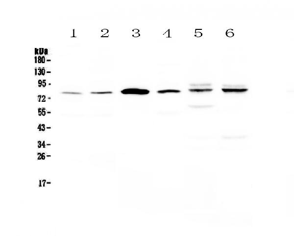
Figure 1. Western blot analysis of CEP68 using anti-CEP68 antibody (A01704-1). Electrophoresis was performed on a 5-20% SDS-PAGE gel at 70V (Stacking gel) / 90V (Resolving gel) for 2-3 hours. The sample well of each lane was loaded with 50ug of sample under reducing conditions. Lane 1: human Hela whole cell lysates, Lane 2: human COLO-320 whole cell lysates, Lane 3: human SK-OV-3 whole cell lysates, Lane 4: human Jurkat whole cell lysates, Lane 5: rat heart tissue lysates,Lane 6: mouse heart tissue lysates. After Electrophoresis, proteins were transferred to a Nitrocellulose membrane at 150mA for 50-90 minutes. Blocked the membrane with 5% Non-fat Milk/ TBS for 1.5 hour at RT. The membrane was incubated with rabbit anti-CEP68 antigen affinity purified polyclonal antibody (Catalog # A01704-1) at 0.5 I?g/mL overnight at 4A°C, then washed with TBS-0.1%Tween 3 times with 5 minutes each and probed with a goat anti-rabbit IgG-HRP secondary antibody at a dilution of 1:10000 for 1.5 hour at RT. The signal is developed using an Enhanced Chemiluminescent detection (ECL) kit (Catalog # EK1002) with Tanon 5200 system. A specific band was detected for CEP68 at approximately 81KD. The expected band size for CEP68 is at 81KD.
|
|
|
|
Figure 1. Western blot analysis of CEP68 using anti-CEP68 antibody (A01704-1). Electrophoresis was performed on a 5-20% SDS-PAGE gel at 70V (Stacking gel) / 90V (Resolving gel) for 2-3 hours. The sample well of each lane was loaded with 50ug of sample under reducing conditions. Lane 1: human Hela whole cell lysates, Lane 2: human COLO-320 whole cell lysates, Lane 3: human SK-OV-3 whole cell lysates, Lane 4: human Jurkat whole cell lysates, Lane 5: rat heart tissue lysates,Lane 6: mouse heart tissue lysates. After Electrophoresis, proteins were transferred to a Nitrocellulose membrane at 150mA for 50-90 minutes. Blocked the membrane with 5% Non-fat Milk/ TBS for 1.5 hour at RT. The membrane was incubated with rabbit anti-CEP68 antigen affinity purified polyclonal antibody (Catalog # A01704-1) at 0.5 I?g/mL overnight at 4A°C, then washed with TBS-0.1%Tween 3 times with 5 minutes each and probed with a goat anti-rabbit IgG-HRP secondary antibody at a dilution of 1:10000 for 1.5 hour at RT. The signal is developed using an Enhanced Chemiluminescent detection (ECL) kit (Catalog # EK1002) with Tanon 5200 system. A specific band was detected for CEP68 at approximately 81KD. The expected band size for CEP68 is at 81KD.
|
|
|
| メーカー |
品番 |
包装 |
|
BBT
|
A01704-1
|
100 UG
|
※表示価格について
| 当社在庫 |
なし
|
| 納期目安 |
1週間程度
|
| 法規制 |
毒
|
| 保存温度 |
-20℃
|
|

