| 別品名 |
SUMO-activating enzyme subunit 2; Anthracycline-associated resistance ARX; Ubiquitin-like 1-activating enzyme E1B; Ubiquitin-like modifier-activating enzyme 2; UBA2; SAE2; UBLE1B; HRIHFB2115
|
| 抗原部位 |
a.a.449-564
|
| 種由来 |
Human
|
| 標識物 |
Unlabeled
|
| 精製度 |
Affinity Purified
|
| 適用 |
Western Blot
IHC paraffin embedding section
Immunocytochemistry (cell)
IHC frozen section
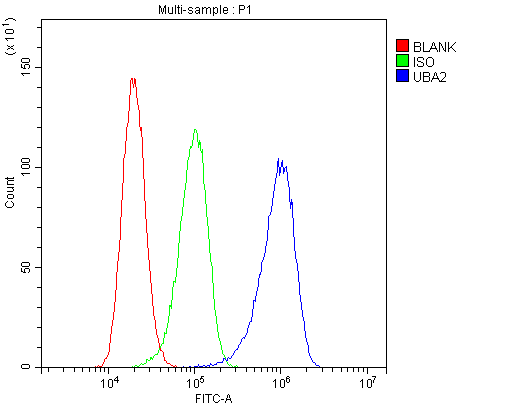
Flow Cytometry
Enzyme Linked Immunosorbent Assay
Immuno Fluorescence
|
| 免疫動物 |
Rabbit
|
| 抗体クラス |
IgG
|
| 交差種 |
Human
Mouse
Rat
|
| GENE ID |
10054
|
| Accession No.(Gene/Protein) |
Q9UBT2
|
| Gene Symbol |
UBA2
|
| 形状 |
凍結乾燥品
|
| 参考文献 |
1. ''Entrez Gene: ubiquitin-like modifier activating enzyme 2''.
2. Gong L, Li B, Millas S, Yeh ET (April 1999). ''Molecular cloning and characterization of human AOS1 and UBA2, components of the sentrin-activating enzyme complex''. FEBS Lett. 448 (1): 185-9.
3. Desterro JM, Rodriguez MS, Kemp GD, Hay RT (April 1999). ''Identification of the enzyme required for activation of the small ubiquitin-like protein SUMO-1''. J. Biol. Chem. 274 (15): 10618-24.
|
|
※サムネイル画像をクリックすると拡大画像が表示されます。
Figure 1. Western blot analysis of SAE2/UBA2 using anti-SAE2/UBA2 antibody (A03816-2). Electrophoresis was performed on a 5-20% SDS-PAGE gel at 70V (Stacking gel) / 90V (Resolving gel) for 2-3 hours. The sample well of each lane was loaded with 50ug of sample under reducing conditions. Lane 1: human Hela whole cell lysates,Lane 2: human placenta tissue lysates,Lane 3: human MCF-7 whole cell lysates,Lane 4: human A549 whole cell lysates,Lane 5: human SK-OV-3 whole cell lysates,Lane 6: human 22RV1 whole cell lysates,Lane 7: human A431 whole cell lysates,Lane 8: human COLO-320 whole cell lysates. After Electrophoresis, proteins were transferred to a Nitrocellulose membrane at 150mA for 50-90 minutes. Blocked the membrane with 5% Non-fat Milk/ TBS for 1.5 hour at RT. The membrane was incubated with rabbit anti-SAE2/UBA2 antigen affinity purified polyclonal antibody (Catalog # A03816-2) at 0.5 I?g/mL overnight at 4A°C, then washed with TBS-0.1%Tween 3 times with 5 minutes each and probed with a goat anti-rabbit IgG-HRP secondary antibody at a dilution of 1:10000 for 1.5 hour at RT. The signal is developed using an Enhanced Chemiluminescent detection (ECL) kit (Catalog # EK1002) with Tanon 5200 system. A specific band was detected for SAE2/UBA2 at approximately 90KD. The expected band size for SAE2/UBA2 is at 71KD.
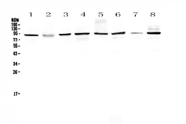
Figure 2. Western blot analysis of SAE2/UBA2 using anti-SAE2/UBA2 antibody (A03816-2). Electrophoresis was performed on a 5-20% SDS-PAGE gel at 70V (Stacking gel) / 90V (Resolving gel) for 2-3 hours. The sample well of each lane was loaded with 50ug of sample under reducing conditions. Lane 1: rat brain tissue lysates,Lane 2: rat lung tissue lysates,Lane 3: rat liver tissue lysates,Lane 4: mouse brain tissue lysates,Lane 5: mouse lung tissue lysates,Lane 6: mouse liver tissue lysates,Lane 7: mouse testis tissue lysates. After Electrophoresis, proteins were transferred to a Nitrocellulose membrane at 150mA for 50-90 minutes. Blocked the membrane with 5% Non-fat Milk/ TBS for 1.5 hour at RT. The membrane was incubated with rabbit anti-SAE2/UBA2 antigen affinity purified polyclonal antibody (Catalog # A03816-2) at 0.5 I?g/mL overnight at 4A°C, then washed with TBS-0.1%Tween 3 times with 5 minutes each and probed with a goat anti-rabbit IgG-HRP secondary antibody at a dilution of 1:10000 for 1.5 hour at RT. The signal is developed using an Enhanced Chemiluminescent detection (ECL) kit (Catalog # EK1002) with Tanon 5200 system. A specific band was detected for SAE2/UBA2 at approximately 90KD. The expected band size for SAE2/UBA2 is at 71KD.
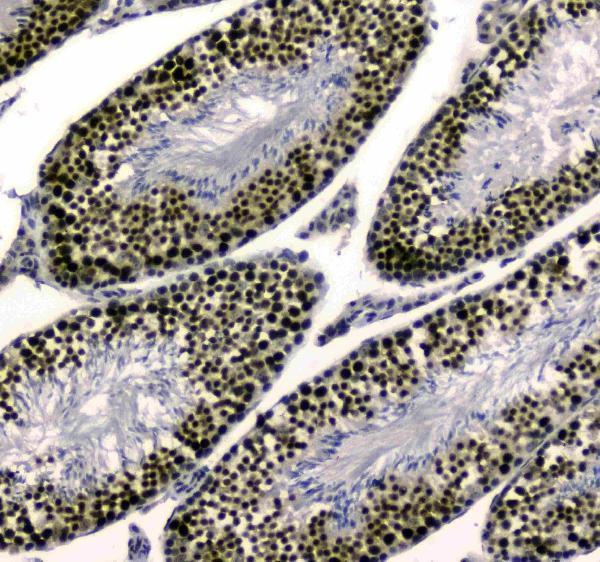
Figure 3. IHC analysis of SAE2/UBA2 using anti-SAE2/UBA2 antibody (A03816-2).SAE2/UBA2 was detected in paraffin-embedded section of human colon cancer tissue. Heat mediated antigen retrieval was performed in citrate buffer (pH6, epitope retrieval solution) for 20 mins. The tissue section was blocked with 10% goat serum. The tissue section was then incubated with 1I?g/ml rabbit anti-SAE2/UBA2 Antibody (A03816-2) overnight at 4A°C. Biotinylated goat anti-rabbit IgG was used as secondary antibody and incubated for 30 minutes at 37A°C. The tissue section was developed using Strepavidin-Biotin-Complex (SABC)(Catalog # SA1022) with DAB as the chromogen.
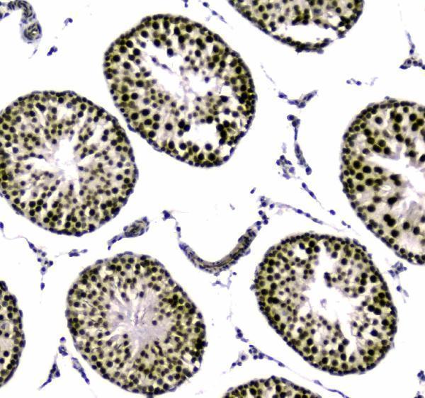
Figure 4. IHC analysis of SAE2/UBA2 using anti-SAE2/UBA2 antibody (A03816-2).SAE2/UBA2 was detected in paraffin-embedded section of human mammary cancer tissue. Heat mediated antigen retrieval was performed in citrate buffer (pH6, epitope retrieval solution) for 20 mins. The tissue section was blocked with 10% goat serum. The tissue section was then incubated with 1I?g/ml rabbit anti-SAE2/UBA2 Antibody (A03816-2) overnight at 4A°C. Biotinylated goat anti-rabbit IgG was used as secondary antibody and incubated for 30 minutes at 37A°C. The tissue section was developed using Strepavidin-Biotin-Complex (SABC)(Catalog # SA1022) with DAB as the chromogen.
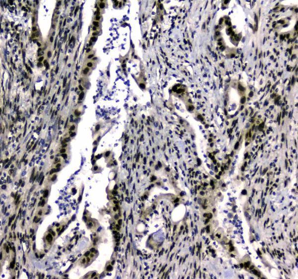
Figure 5. IHC analysis of SAE2/UBA2 using anti-SAE2/UBA2 antibody (A03816-2).SAE2/UBA2 was detected in paraffin-embedded section of rat testis tissue. Heat mediated antigen retrieval was performed in citrate buffer (pH6, epitope retrieval solution) for 20 mins. The tissue section was blocked with 10% goat serum. The tissue section was then incubated with 1I?g/ml rabbit anti-SAE2/UBA2 Antibody (A03816-2) overnight at 4A°C. Biotinylated goat anti-rabbit IgG was used as secondary antibody and incubated for 30 minutes at 37A°C. The tissue section was developed using Strepavidin-Biotin-Complex (SABC)(Catalog # SA1022) with DAB as the chromogen.
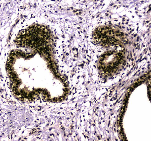
Figure 6. IHC analysis of SAE2/UBA2 using anti-SAE2/UBA2 antibody (A03816-2).SAE2/UBA2 was detected in paraffin-embedded section of mouse testis tissue. Heat mediated antigen retrieval was performed in citrate buffer (pH6, epitope retrieval solution) for 20 mins. The tissue section was blocked with 10% goat serum. The tissue section was then incubated with 1I?g/ml rabbit anti-SAE2/UBA2 Antibody (A03816-2) overnight at 4A°C. Biotinylated goat anti-rabbit IgG was used as secondary antibody and incubated for 30 minutes at 37A°C. The tissue section was developed using Strepavidin-Biotin-Complex (SABC)(Catalog # SA1022) with DAB as the chromogen.
|
|
|
|
Figure 1. Western blot analysis of SAE2/UBA2 using anti-SAE2/UBA2 antibody (A03816-2). Electrophoresis was performed on a 5-20% SDS-PAGE gel at 70V (Stacking gel) / 90V (Resolving gel) for 2-3 hours. The sample well of each lane was loaded with 50ug of sample under reducing conditions. Lane 1: human Hela whole cell lysates,Lane 2: human placenta tissue lysates,Lane 3: human MCF-7 whole cell lysates,Lane 4: human A549 whole cell lysates,Lane 5: human SK-OV-3 whole cell lysates,Lane 6: human 22RV1 whole cell lysates,Lane 7: human A431 whole cell lysates,Lane 8: human COLO-320 whole cell lysates. After Electrophoresis, proteins were transferred to a Nitrocellulose membrane at 150mA for 50-90 minutes. Blocked the membrane with 5% Non-fat Milk/ TBS for 1.5 hour at RT. The membrane was incubated with rabbit anti-SAE2/UBA2 antigen affinity purified polyclonal antibody (Catalog # A03816-2) at 0.5 I?g/mL overnight at 4A°C, then washed with TBS-0.1%Tween 3 times with 5 minutes each and probed with a goat anti-rabbit IgG-HRP secondary antibody at a dilution of 1:10000 for 1.5 hour at RT. The signal is developed using an Enhanced Chemiluminescent detection (ECL) kit (Catalog # EK1002) with Tanon 5200 system. A specific band was detected for SAE2/UBA2 at approximately 90KD. The expected band size for SAE2/UBA2 is at 71KD.
|
|
|
| メーカー |
品番 |
包装 |
|
BBT
|
A03816-2
|
100 UG
|
※表示価格について
| 当社在庫 |
なし
|
| 納期目安 |
1週間程度
|
| 法規制 |
毒
|
| 保存温度 |
-20℃
|
|