| 別品名 |
N-acetylgalactosamine-6-sulfatase; Chondroitinsulfatase; Chondroitinase; Galactose-6-sulfate sulfatase; GalN6S; N-acetylgalactosamine-6-sulfate sulfatase; GalNAc6S sulfatase; GALNS
|
| 抗原部位 |
a.a.181-289
|
| 種由来 |
Human
|
| 標識物 |
Unlabeled
|
| 精製度 |
Affinity Purified
|
| 適用 |
Western Blot
IHC paraffin embedding section
Enzyme Linked Immunosorbent Assay
|
| 免疫動物 |
Rabbit
|
| 抗体クラス |
IgG
|
| 交差種 |
Human
Mouse
Rat
|
| GENE ID |
2588
|
| Accession No.(Gene/Protein) |
P34059
|
| Gene Symbol |
GALNS
|
| 分子量 |
58026 MW
|
| 形状 |
凍結乾燥品
|
| 参考文献 |
1. ''Entrez Gene: GALNS galactosamine (N-acetyl)-6-sulfate sulfatase (Morquio syndrome, mucopolysaccharidosis type IVA)''.
2. Tomatsu S, Fukuda S, Masue M, Sukegawa K, Fukao T, Yamagishi A, Hori T, Iwata H, Ogawa T, Nakashima Y, et al. (Jan 1992). ''Morquio disease: isolation, characterization and expression of full-length cDNA for human N-acetylgalactosamine-6-sulfate sulfatase''. Biochem Biophys Res Commun. 181 (2): 677-83.
|
|
※サムネイル画像をクリックすると拡大画像が表示されます。
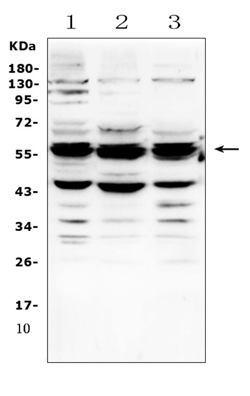
Western blot analysis of GALNS using anti-GALNS antibody (A02954-1).
Electrophoresis was performed on a 5-20% SDS-PAGE gel at 70V (Stacking gel) / 90V (Resolving gel) for 2-3 hours. The sample well of each lane was loaded with 50ug of sample under reducing conditions.
Lane 1: rat small intestine tissue lysates.
After Electrophoresis, proteins were transferred to a Nitrocellulose membrane at 150mA for 50-90 minutes. Blocked the membrane with 5% Non-fat Milk/ TBS for 1.5 hour at RT. The membrane was incubated with rabbit anti-GALNS antigen affinity purified polyclonal antibody (Catalog # A02954-1) at 0.5 I?g/mL overnight at 4A°C, then washed with TBS-0.1%Tween 3 times with 5 minutes each and probed with a goat anti-rabbit IgG-HRP secondary antibody at a dilution of 1:10000 for 1.5 hour at RT. The signal is developed using an Enhanced Chemiluminescent detection (ECL) kit (Catalog # EK1002) with Tanon 5200 system. A specific band was detected for GALNS at approximately 58KD. The expected band size for GALNS is at 58KD.
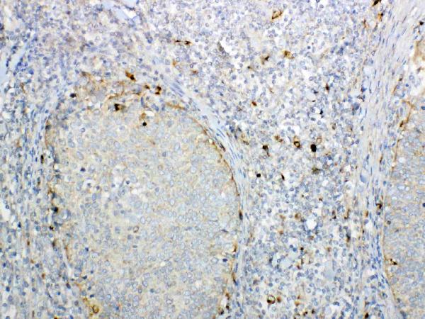
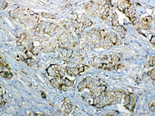
IHC analysis of GALNS using anti-GALNS antibody (A02954-1).
GALNS was detected in paraffin-embedded section of human lung cancer tissue. Heat mediated antigen retrieval was performed in citrate buffer (pH6, epitope retrieval solution) for 20 mins. The tissue section was blocked with 10% goat serum. The tissue section was then incubated with 1I?g/ml rabbit anti-GALNS Antibody (A02954-1) overnight at 4A°C. Biotinylated goat anti-rabbit IgG was used as secondary antibody and incubated for 30 minutes at 37A°C. The tissue section was developed using Strepavidin-Biotin-Complex (SABC)(Catalog # SA1022) with DAB as the chromogen.
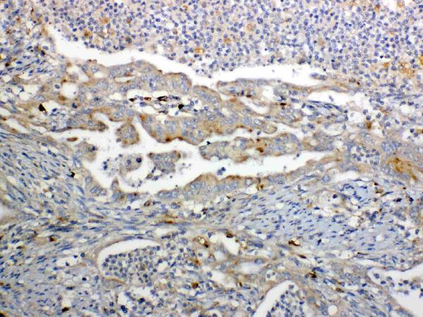
IHC analysis of GALNS using anti-GALNS antibody (A02954-1).
GALNS was detected in paraffin-embedded section of human colon cancer tissue. Heat mediated antigen retrieval was performed in citrate buffer (pH6, epitope retrieval solution) for 20 mins. The tissue section was blocked with 10% goat serum. The tissue section was then incubated with 1I?g/ml rabbit anti-GALNS Antibody (A02954-1) overnight at 4A°C. Biotinylated goat anti-rabbit IgG was used as secondary antibody and incubated for 30 minutes at 37A°C. The tissue section was developed using Strepavidin-Biotin-Complex (SABC)(Catalog # SA1022) with DAB as the chromogen.
IHC analysis of GALNS using anti-GALNS antibody (A02954-1).
GALNS was detected in paraffin-embedded section of human mammary cancer tissue. Heat mediated antigen retrieval was performed in citrate buffer (pH6, epitope retrieval solution) for 20 mins. The tissue section was blocked with 10% goat serum. The tissue section was then incubated with 1I?g/ml rabbit anti-GALNS Antibody (A02954-1) overnight at 4A°C. Biotinylated goat anti-rabbit IgG was used as secondary antibody and incubated for 30 minutes at 37A°C. The tissue section was developed using Strepavidin-Biotin-Complex (SABC)(Catalog # SA1022) with DAB as the chromogen.
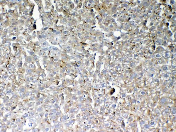
IHC analysis of GALNS using anti-GALNS antibody (A02954-1).
GALNS was detected in paraffin-embedded section of mouse liver tissue. Heat mediated antigen retrieval was performed in citrate buffer (pH6, epitope retrieval solution) for 20 mins. The tissue section was blocked with 10% goat serum. The tissue section was then incubated with 1I?g/ml rabbit anti-GALNS Antibody (A02954-1) overnight at 4A°C. Biotinylated goat anti-rabbit IgG was used as secondary antibody and incubated for 30 minutes at 37A°C. The tissue section was developed using Strepavidin-Biotin-Complex (SABC)(Catalog # SA1022) with DAB as the chromogen.
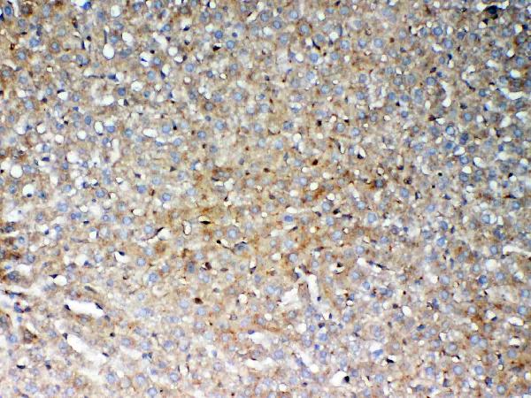
IHC analysis of GALNS using anti-GALNS antibody (A02954-1).
GALNS was detected in paraffin-embedded section of rat liver tissue. Heat mediated antigen retrieval was performed in citrate buffer (pH6, epitope retrieval solution) for 20 mins. The tissue section was blocked with 10% goat serum. The tissue section was then incubated with 1I?g/ml rabbit anti-GALNS Antibody (A02954-1) overnight at 4A°C. Biotinylated goat anti-rabbit IgG was used as secondary antibody and incubated for 30 minutes at 37A°C. The tissue section was developed using Strepavidin-Biotin-Complex (SABC)(Catalog # SA1022) with DAB as the chromogen.
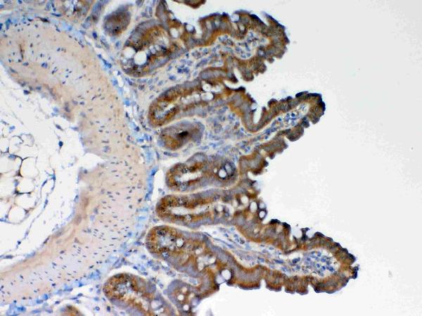
IHC analysis of GALNS using anti-GALNS antibody (A02954-1).
GALNS was detected in paraffin-embedded section of rat small intestine tissue. Heat mediated antigen retrieval was performed in citrate buffer (pH6, epitope retrieval solution) for 20 mins. The tissue section was blocked with 10% goat serum. The tissue section was then incubated with 1I?g/ml rabbit anti-GALNS Antibody (A02954-1) overnight at 4A°C. Biotinylated goat anti-rabbit IgG was used as secondary antibody and incubated for 30 minutes at 37A°C. The tissue section was developed using Strepavidin-Biotin-Complex (SABC)(Catalog # SA1022) with DAB as the chromogen.
|
|
|
|
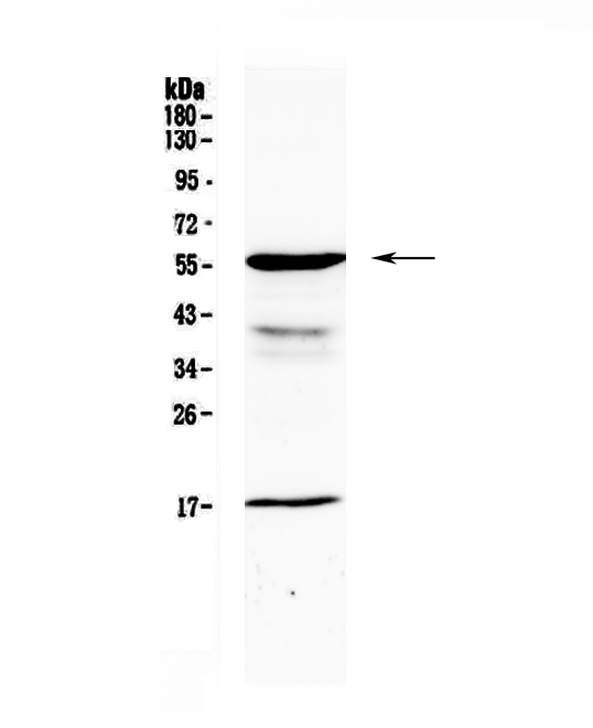
Western blot analysis of GALNS using anti-GALNS antibody (A02954-1).
Electrophoresis was performed on a 5-20% SDS-PAGE gel at 70V (Stacking gel) / 90V (Resolving gel) for 2-3 hours. The sample well of each lane was loaded with 50ug of sample under reducing conditions.
Lane 1: rat small intestine tissue lysates.
After Electrophoresis, proteins were transferred to a Nitrocellulose membrane at 150mA for 50-90 minutes. Blocked the membrane with 5% Non-fat Milk/ TBS for 1.5 hour at RT. The membrane was incubated with rabbit anti-GALNS antigen affinity purified polyclonal antibody (Catalog # A02954-1) at 0.5 I?g/mL overnight at 4A°C, then washed with TBS-0.1%Tween 3 times with 5 minutes each and probed with a goat anti-rabbit IgG-HRP secondary antibody at a dilution of 1:10000 for 1.5 hour at RT. The signal is developed using an Enhanced Chemiluminescent detection (ECL) kit (Catalog # EK1002) with Tanon 5200 system. A specific band was detected for GALNS at approximately 58KD. The expected band size for GALNS is at 58KD.
|
|
|
| メーカー |
品番 |
包装 |
|
BBT
|
A02954-1
|
100 UG
|
※表示価格について
| 当社在庫 |
なし
|
| 納期目安 |
1週間程度
|
| 法規制 |
毒
|
| 保存温度 |
-20℃
|
|