

| 別品名 |
Dynamin-1; DNM1; DNM
|
| 抗原部位 |
a.a.616-667
|
| 種由来 |
Human
|
| 標識物 |
Unlabeled
|
| 適用 |
Western Blot
Enzyme Linked Immunosorbent Assay
Immunohistochemistry
Immunocytochemistry (cell)
Flow Cytometry
|
| 免疫動物 |
Rabbit
|
| 抗体クラス |
IgG
|
| 交差種 |
Human
Mouse
Rat
|
| GENE ID |
1759
|
| Accession No.(Gene/Protein) |
Q05193
|
| Gene Symbol |
DNM1
|
| 分子量 |
97 kDa
|
| 形状 |
凍結乾燥品
|
| 参考文献 |
1. Dhindsa, R. S., Bradrick, S. S., Yao, X., Heinzen, E. L., Petrovski, S., Krueger, B. J., Johnson, M. R., Frankel, W. N., Petrou, S., Boumil, R. M., Goldstein, D. B. Epileptic encephalopathy-causing mutations in DNM1 impair synaptic vesicle endocytosis. Neurol. Genet. 1: e4, 2015.
2. EuroEPINOMICS-RES Consortium, Epilepsy Phenome/Genome Project, Epi4K Consortium. De novo mutations in synaptic transmission genes including DNM1 cause epileptic encephalopathies. Am. J. Hum. Genet. 95: 360-370, 2014.
|
|
※サムネイル画像をクリックすると拡大画像が表示されます。
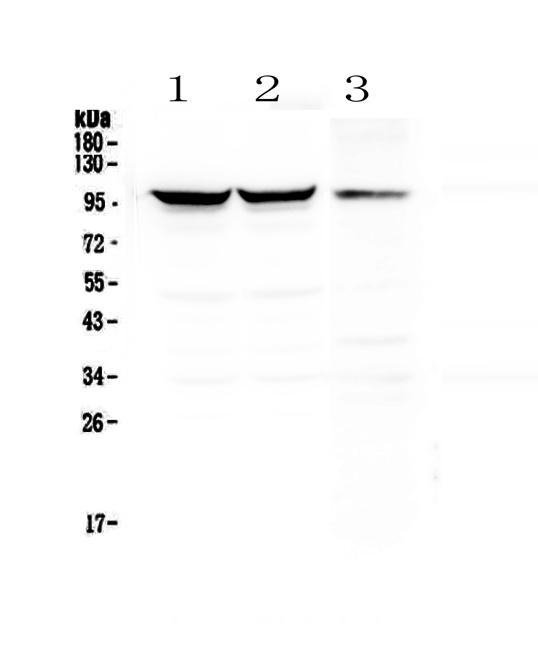
Figure 1. Western blot analysis of Dynamin 1 using anti-Dynamin 1 antibody (A02536-2). Electrophoresis was performed on a 5-20% SDS-PAGE gel at 70V (Stacking gel) / 90V (Resolving gel) for 2-3 hours. The sample well of each lane was loaded with 50ug of sample under reducing conditions. Lane 1: rat brain tissue lysates,Lane 2: mouse brain tissue lysates,Lane 3: mouse NIH3T3 whole cell lysates. After Electrophoresis, proteins were transferred to a Nitrocellulose membrane at 150mA for 50-90 minutes. Blocked the membrane with 5% Non-fat Milk/ TBS for 1.5 hour at RT. The membrane was incubated with rabbit anti-Dynamin 1 antigen affinity purified polyclonal antibody (Catalog # A02536-2) at 0.5 I?g/mL overnight at 4A°C, then washed with TBS-0.1%Tween 3 times with 5 minutes each and probed with a goat anti-rabbit IgG-HRP secondary antibody at a dilution of 1:10000 for 1.5 hour at RT. The signal is developed using an Enhanced Chemiluminescent detection (ECL) kit (Catalog # EK1002) with Tanon 5200 system. A specific band was detected for Dynamin 1 at approximately 97KD. The expected band size for Dynamin 1 is at 97KD.
|
|
|
|
Figure 1. Western blot analysis of Dynamin 1 using anti-Dynamin 1 antibody (A02536-2). Electrophoresis was performed on a 5-20% SDS-PAGE gel at 70V (Stacking gel) / 90V (Resolving gel) for 2-3 hours. The sample well of each lane was loaded with 50ug of sample under reducing conditions. Lane 1: rat brain tissue lysates,Lane 2: mouse brain tissue lysates,Lane 3: mouse NIH3T3 whole cell lysates. After Electrophoresis, proteins were transferred to a Nitrocellulose membrane at 150mA for 50-90 minutes. Blocked the membrane with 5% Non-fat Milk/ TBS for 1.5 hour at RT. The membrane was incubated with rabbit anti-Dynamin 1 antigen affinity purified polyclonal antibody (Catalog # A02536-2) at 0.5 I?g/mL overnight at 4A°C, then washed with TBS-0.1%Tween 3 times with 5 minutes each and probed with a goat anti-rabbit IgG-HRP secondary antibody at a dilution of 1:10000 for 1.5 hour at RT. The signal is developed using an Enhanced Chemiluminescent detection (ECL) kit (Catalog # EK1002) with Tanon 5200 system. A specific band was detected for Dynamin 1 at approximately 97KD. The expected band size for Dynamin 1 is at 97KD.
|
|
|
| メーカー |
品番 |
包装 |
|
BBT
|
A02536-2
|
100 UG
|
※表示価格について
| 当社在庫 |
なし
|
| 納期目安 |
1週間程度
|
| 法規制 |
毒
|
| 保存温度 |
-20℃
|
|

