

| 別品名 |
Deoxycytidine kinase; dCK; DCK;
|
| 抗原部位 |
a.a.17-260
|
| 種由来 |
Human
|
| 標識物 |
Unlabeled
|
| 精製度 |
Affinity Purified
|
| 適用 |
Western Blot
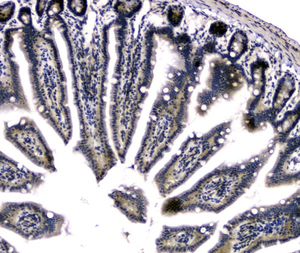
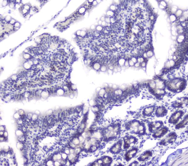
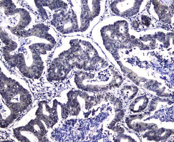
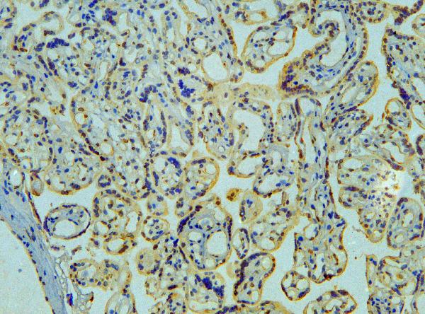
IHC paraffin embedding section
IHC frozen section
Immunocytochemistry (cell)
Flow Cytometry
Enzyme Linked Immunosorbent Assay
|
| 免疫動物 |
Rabbit
|
| 抗体クラス |
IgG
|
| 交差種 |
Human
Mouse
Rat
|
| GENE ID |
1633
|
| Accession No.(Gene/Protein) |
P27707
|
| Gene Symbol |
DCK
|
| 形状 |
凍結乾燥品
|
| 参考文献 |
1. Chottiner, E. G., Shewach, D. S., Datta, N. S., Ashcraft, E., Gribbin, D., Ginsburg, D., Fox, I. H., Mitchell, B. S. Cloning and expression of human deoxycytidine kinase cDNA. Proc. Nat. Acad. Sci. 88: 1531-1535, 1991.
2. Stegmann, A. P. A., Honders, M. W., Bolk, M. W. J., Wessels, J., Willemze, R., Landegent, J. E.Assignment of the human deoxycytidine kinase (DCK) gene to chromosome 4 band q13.3-q21.1. Genomics 17: 528-529, 1993.
|
|
※サムネイル画像をクリックすると拡大画像が表示されます。
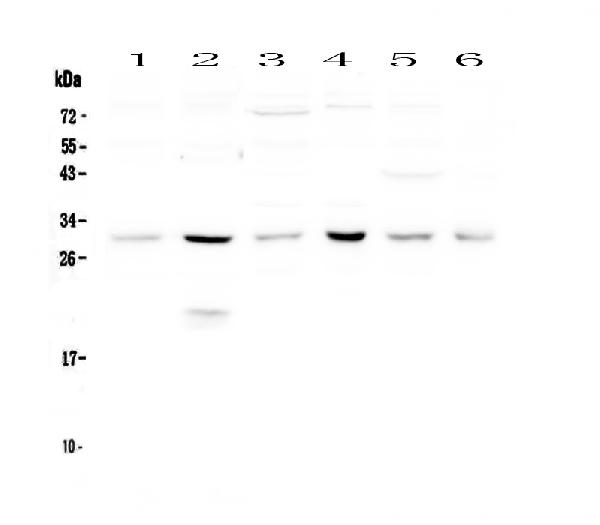
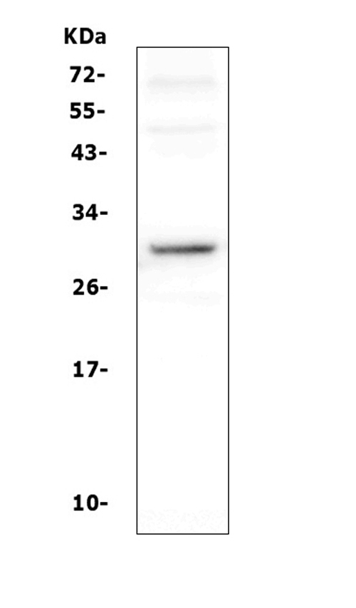
Figure 1. Western blot analysis of DCK using anti-DCK antibody (A01655-1). Electrophoresis was performed on a 5-20% SDS-PAGE gel at 70V (Stacking gel) / 90V (Resolving gel) for 2-3 hours. The sample well of each lane was loaded with 50ug of sample under reducing conditions. Lane 1: mouse spleen tissue lysates,Lane 2: mouse thymus tissue lysates,Lane 3: human Hela whole cell lysates,Lane 4: human U-87MG whole cell lysates,Lane 5: human MCF-7 whole cell lysates,Lane 6: human U20S whole cell lysates. After Electrophoresis, proteins were transferred to a Nitrocellulose membrane at 150mA for 50-90 minutes. Blocked the membrane with 5% Non-fat Milk/ TBS for 1.5 hour at RT. The membrane was incubated with rabbit anti-DCK antigen affinity purified polyclonal antibody (Catalog # A01655-1) at 0.5 I?g/mL overnight at 4A°C, then washed with TBS-0.1%Tween 3 times with 5 minutes each and probed with a goat anti-rabbit IgG-HRP secondary antibody at a dilution of 1:10000 for 1.5 hour at RT. The signal is developed using an Enhanced Chemiluminescent detection (ECL) kit (Catalog # EK1002) with Tanon 5200 system. A specific band was detected for DCK at approximately 30KD. The expected band size for DCK is at 30KD.
|
|
|
|
Figure 1. Western blot analysis of DCK using anti-DCK antibody (A01655-1). Electrophoresis was performed on a 5-20% SDS-PAGE gel at 70V (Stacking gel) / 90V (Resolving gel) for 2-3 hours. The sample well of each lane was loaded with 50ug of sample under reducing conditions. Lane 1: mouse spleen tissue lysates,Lane 2: mouse thymus tissue lysates,Lane 3: human Hela whole cell lysates,Lane 4: human U-87MG whole cell lysates,Lane 5: human MCF-7 whole cell lysates,Lane 6: human U20S whole cell lysates. After Electrophoresis, proteins were transferred to a Nitrocellulose membrane at 150mA for 50-90 minutes. Blocked the membrane with 5% Non-fat Milk/ TBS for 1.5 hour at RT. The membrane was incubated with rabbit anti-DCK antigen affinity purified polyclonal antibody (Catalog # A01655-1) at 0.5 I?g/mL overnight at 4A°C, then washed with TBS-0.1%Tween 3 times with 5 minutes each and probed with a goat anti-rabbit IgG-HRP secondary antibody at a dilution of 1:10000 for 1.5 hour at RT. The signal is developed using an Enhanced Chemiluminescent detection (ECL) kit (Catalog # EK1002) with Tanon 5200 system. A specific band was detected for DCK at approximately 30KD. The expected band size for DCK is at 30KD.
|
|
|
| メーカー |
品番 |
包装 |
|
BBT
|
A01655-1
|
100 UG
|
※表示価格について
| 当社在庫 |
なし
|
| 納期目安 |
1週間程度
|
| 法規制 |
毒
|
| 保存温度 |
-20℃
|
|

