

| 別品名 |
Retinol-binding protein 4; Plasma retinol-binding protein; PRBP; RBP; Rbp4;
|
| 抗原部位 |
a.a.19-201
|
| 種由来 |
Mouse
|
| 標識物 |
Unlabeled
|
| 精製度 |
Affinity Purified
|
| 適用 |
Western Blot
Enzyme Linked Immunosorbent Assay
Immunohistochemistry
|
| 免疫動物 |
Rabbit
|
| 抗体クラス |
IgG
|
| 交差種 |
Human
Mouse
Rat
|
| GENE ID |
19662
|
| Accession No.(Gene/Protein) |
Q00724
|
| Gene Symbol |
RBP4
|
| 分子量 |
23 kDa
|
| 形状 |
凍結乾燥品
|
| 参考文献 |
1. Cubedo J, et al. Retinol-binding protein 4 levels and susceptibility to ischaemic events in men. Eur J Clin Invest, 2014.
2. Li F, et al. Retinol-binding protein 4 as a novel risk factor for cardiovascular disease in patients with coronary artery disease and hyperinsulinemia. Am J Med Sci, 2014 Dec.
3. Saucedo R, et al. RBP4 gene variants are associated with insulin resistance in women with previous gestational diabetes. Dis Markers, 2014.
|
|
※サムネイル画像をクリックすると拡大画像が表示されます。
Western blot analysis of RBP4 using anti-RBP4 antibody (A01089).
Electrophoresis was performed on a 5-20% SDS-PAGE gel at 70V (Stacking gel) / 90V (Resolving gel) for 2-3 hours. The sample well of each lane was loaded with 50ug of sample under reducing conditions.
Lane 1: mouse liver tissue lysates,
Lane 2: rat liver tissue lysates,
Lane 3: human placenta tissue lysates,
Lane 4: human HepG2 whole cell lysates.
After Electrophoresis, proteins were transferred to a Nitrocellulose membrane at 150mA for 50-90 minutes. Blocked the membrane with 5% Non-fat Milk/ TBS for 1.5 hour at RT. The membrane was incubated with rabbit anti-RBP4 antigen affinity purified polyclonal antibody (Catalog # A01089) at 0.5 ug/mL overnight at 4℃, then washed with TBS-0.1%Tween 3 times with 5 minutes each and probed with a goat anti-rabbit IgG-HRP secondary antibody at a dilution of 1:10000 for 1.5 hour at RT. The signal is developed using an Enhanced Chemiluminescent detection (ECL) kit (Catalog # EK1002) with Tanon 5200 system. A specific band was detected for RBP4 at approximately 23KD. The expected band size for RBP4 is at 23KD.
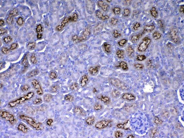
IHC analysis of RBP4 using anti-RBP4 antibody (A01089).
RBP4 was detected in paraffin-embedded section of mouse kidney tissue. Heat mediated antigen retrieval was performed in citrate buffer (pH6, epitope retrieval solution) for 20 mins. The tissue section was blocked with 10% goat serum. The tissue section was then incubated with 1ug/ml rabbit anti-RBP4 Antibody (A01089) overnight at 4℃. Biotinylated goat anti-rabbit IgG was used as secondary antibody and incubated for 30 minutes at 37℃. The tissue section was developed using Strepavidin-Biotin-Complex (SABC)(Catalog # SA1022) with DAB as the chromogen.
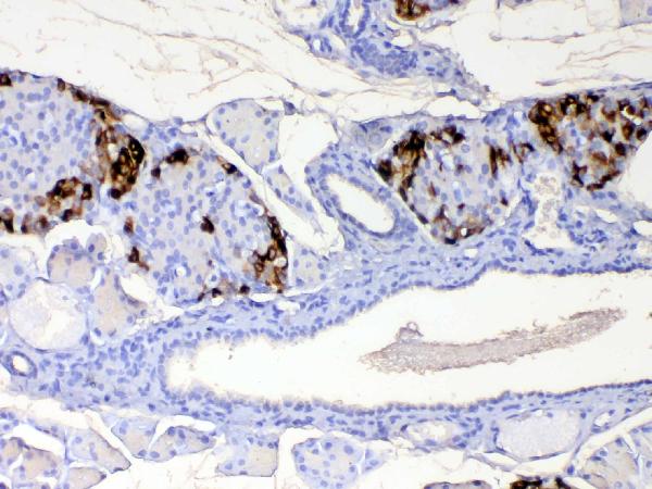
IHC analysis of RBP4 using anti-RBP4 antibody (A01089).
RBP4 was detected in paraffin-embedded section of mouse pancreas tissue. Heat mediated antigen retrieval was performed in citrate buffer (pH6, epitope retrieval solution) for 20 mins. The tissue section was blocked with 10% goat serum. The tissue section was then incubated with 1ug/ml rabbit anti-RBP4 Antibody (A01089) overnight at 4℃. Biotinylated goat anti-rabbit IgG was used as secondary antibody and incubated for 30 minutes at 37℃. The tissue section was developed using Strepavidin-Biotin-Complex (SABC)(Catalog # SA1022) with DAB as the chromogen.
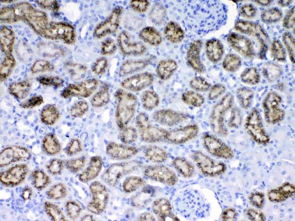
IHC analysis of RBP4 using anti-RBP4 antibody (A01089).
RBP4 was detected in paraffin-embedded section of rat kidney tissue. Heat mediated antigen retrieval was performed in citrate buffer (pH6, epitope retrieval solution) for 20 mins. The tissue section was blocked with 10% goat serum. The tissue section was then incubated with 1ug/ml rabbit anti-RBP4 Antibody (A01089) overnight at 4℃. Biotinylated goat anti-rabbit IgG was used as secondary antibody and incubated for 30 minutes at 37℃. The tissue section was developed using Strepavidin-Biotin-Complex (SABC)(Catalog # SA1022) with DAB as the chromogen.

IHC analysis of RBP4 using anti-RBP4 antibody (A01089).
RBP4 was detected in paraffin-embedded section of rat pancreas tissue. Heat mediated antigen retrieval was performed in citrate buffer (pH6, epitope retrieval solution) for 20 mins. The tissue section was blocked with 10% goat serum. The tissue section was then incubated with 1ug/ml rabbit anti-RBP4 Antibody (A01089) overnight at 4℃. Biotinylated goat anti-rabbit IgG was used as secondary antibody and incubated for 30 minutes at 37℃. The tissue section was developed using Strepavidin-Biotin-Complex (SABC)(Catalog # SA1022) with DAB as the chromogen.

|
|
|
|
Western blot analysis of RBP4 using anti-RBP4 antibody (A01089).
Electrophoresis was performed on a 5-20% SDS-PAGE gel at 70V (Stacking gel) / 90V (Resolving gel) for 2-3 hours. The sample well of each lane was loaded with 50ug of sample under reducing conditions.
Lane 1: mouse liver tissue lysates,
Lane 2: rat liver tissue lysates,
Lane 3: human placenta tissue lysates,
Lane 4: human HepG2 whole cell lysates.
After Electrophoresis, proteins were transferred to a Nitrocellulose membrane at 150mA for 50-90 minutes. Blocked the membrane with 5% Non-fat Milk/ TBS for 1.5 hour at RT. The membrane was incubated with rabbit anti-RBP4 antigen affinity purified polyclonal antibody (Catalog # A01089) at 0.5 ug/mL overnight at 4℃, then washed with TBS-0.1%Tween 3 times with 5 minutes each and probed with a goat anti-rabbit IgG-HRP secondary antibody at a dilution of 1:10000 for 1.5 hour at RT. The signal is developed using an Enhanced Chemiluminescent detection (ECL) kit (Catalog # EK1002) with Tanon 5200 system. A specific band was detected for RBP4 at approximately 23KD. The expected band size for RBP4 is at 23KD.
|
|
|
| メーカー |
品番 |
包装 |
|
BBT
|
A01089
|
100 UG
|
※表示価格について
| 当社在庫 |
なし
|
| 納期目安 |
1週間程度
|
| 法規制 |
毒
|
| 保存温度 |
-20℃
|
|

