|
※サムネイル画像をクリックすると拡大画像が表示されます。
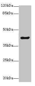
Western blot
All lanes: MEST antibody at 5.78ug/ml + Mouse kidney tissue
Secondary
Goat polyclonal to rabbit IgG at 1/10000 dilution
Predicted band size: 39, 38, 34 kDa
Observed band size: 39 kDa
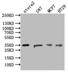
Western Blot
Positive WB detected in: NTERA-2 whole cell lysate, U87 whole cell lysate, MCF7 whole cell lysate, HT29 whole cell lysate
All lanes: MEST antibody at 1:500
Secondary
Goat polyclonal to rabbit IgG at 1/50000 dilution
Predicted band size: 39, 38, 34 kDa
Observed band size: 35 kDa
Immunohistochemistry of paraffin-embedded human pancreatic tissue using CSB-PA704858ESR1HU at dilution of 1:100

Immunohistochemistry of paraffin-embedded human Pancreatic tissue using CSB-PA704858ESR1HU at dilution of 1:50
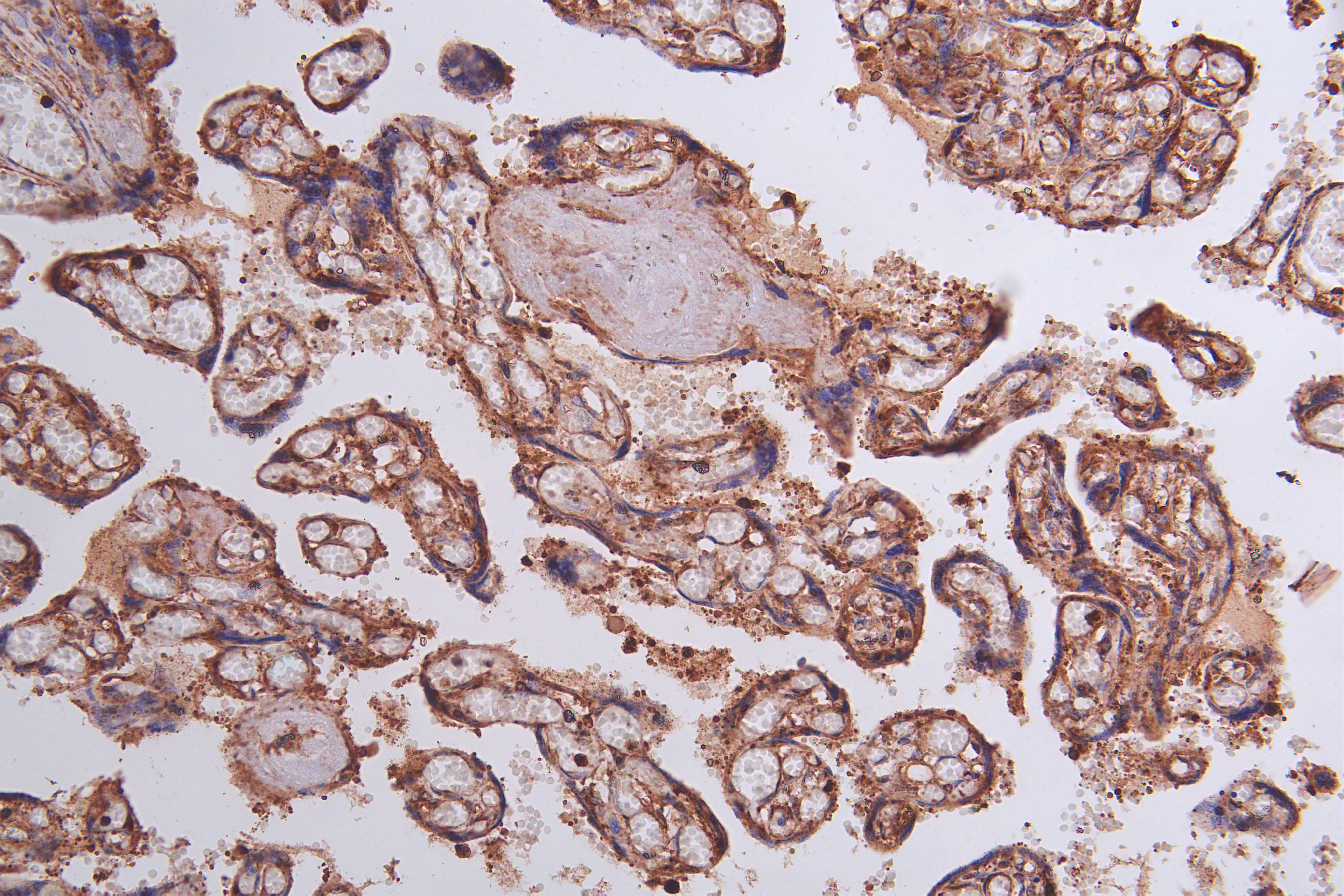
Immunohistochemistry?of?paraffin-embedded?human?Breast cancer?using?CSB-PA704858ESR1HU?at?dilution?of?1:50

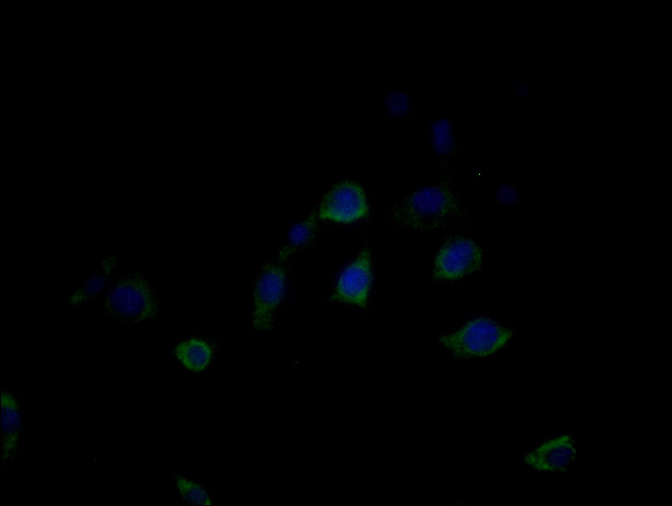
Immunofluorescence staining of MCF-7 cells with CSB-PA704858ESR1HU at 1:25, counter-stained with DAPI. The cells were fixed in 4% formaldehyde, permeabilized using 0.2% Triton X-100 and blocked in 10% normal Goat Serum. The cells were then incubated with the antibody overnight at 4°C. The secondary antibody was Alexa Fluor 488-congugated AffiniPure Goat Anti-Rabbit IgG(H+L).
|

