|
※サムネイル画像をクリックすると拡大画像が表示されます。
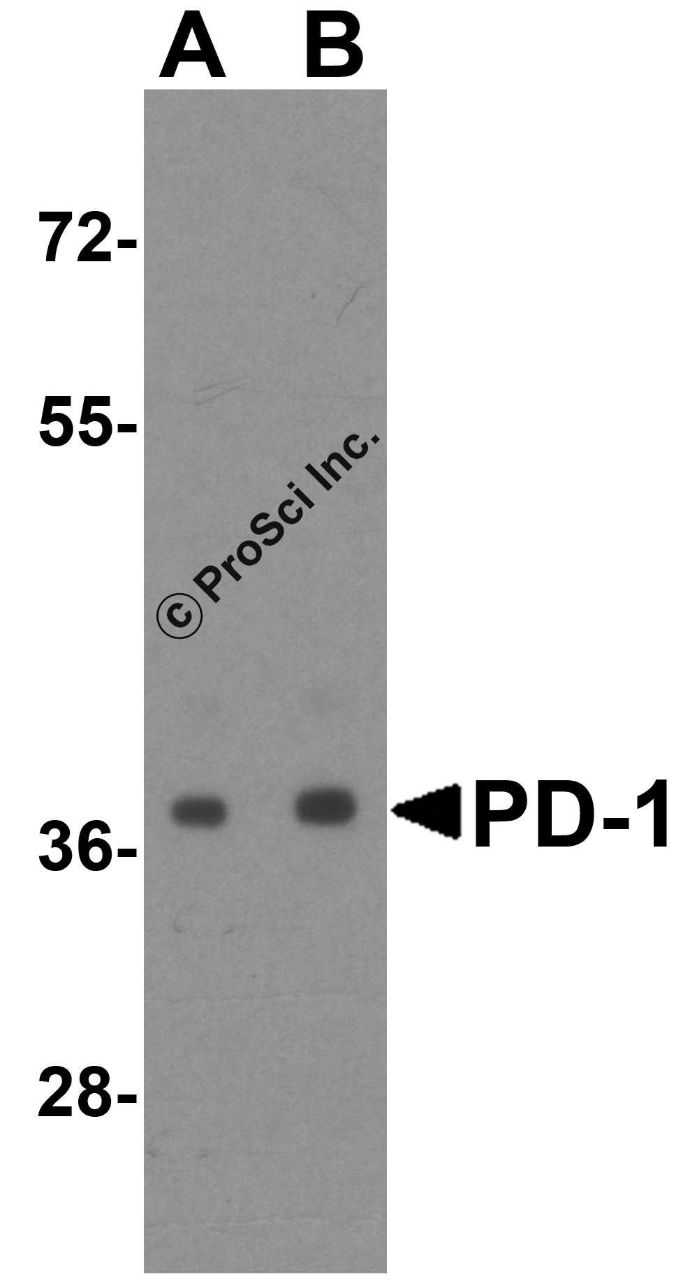
Western blot analysis of PD-1 in transfected 293 cell lysate with PD-1 antibody at (A) 0.5 and (B) 1 μg/mL.
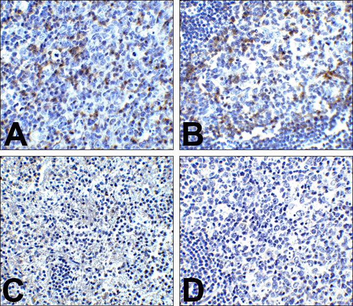
Immunohistochemistry of PD-1 in (A) human tonsil tissue, (B) human lymph node tissue, and (C) human spleen tissue with PD-1 antibody at 5 μg/mL. (D) Immunohistochemistry in human tonsil tissue with control mouse IgG staining at 5 μg/mL.
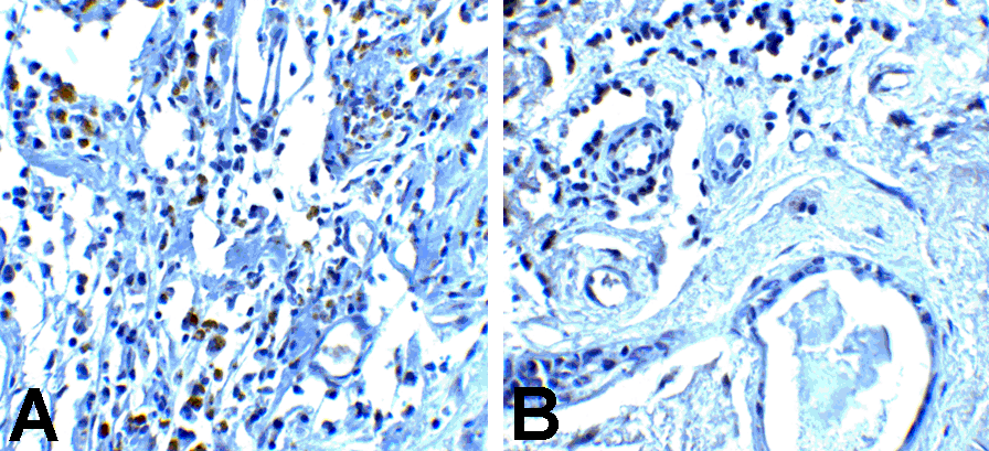
Immunohistochemistry of PD-1 in (A) human breast cancer tissue and (B) human normal breast tissue with PD-1 antibody at 5 μg/mL.
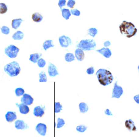
Immunocytochemistry of PD-1 in transfected 293 cells with PD-1 antibody at 5 μg/mL. Lower left: Immunocytochemistry in transfected 293 cells with control mouse IgG antibody at 5 μg/mL.
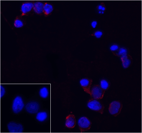
Immunofluorescence of PD-1 in transfected 293 cells with PD-1 antibody at 5 μg/mL. Lower left: Immunofluorescence in transfected 293 cells with control mouse IgG antibody at 5 μg/mL.
Red: PD1 Antibody [8A4] (RF16002)
Blue: DAPI staining
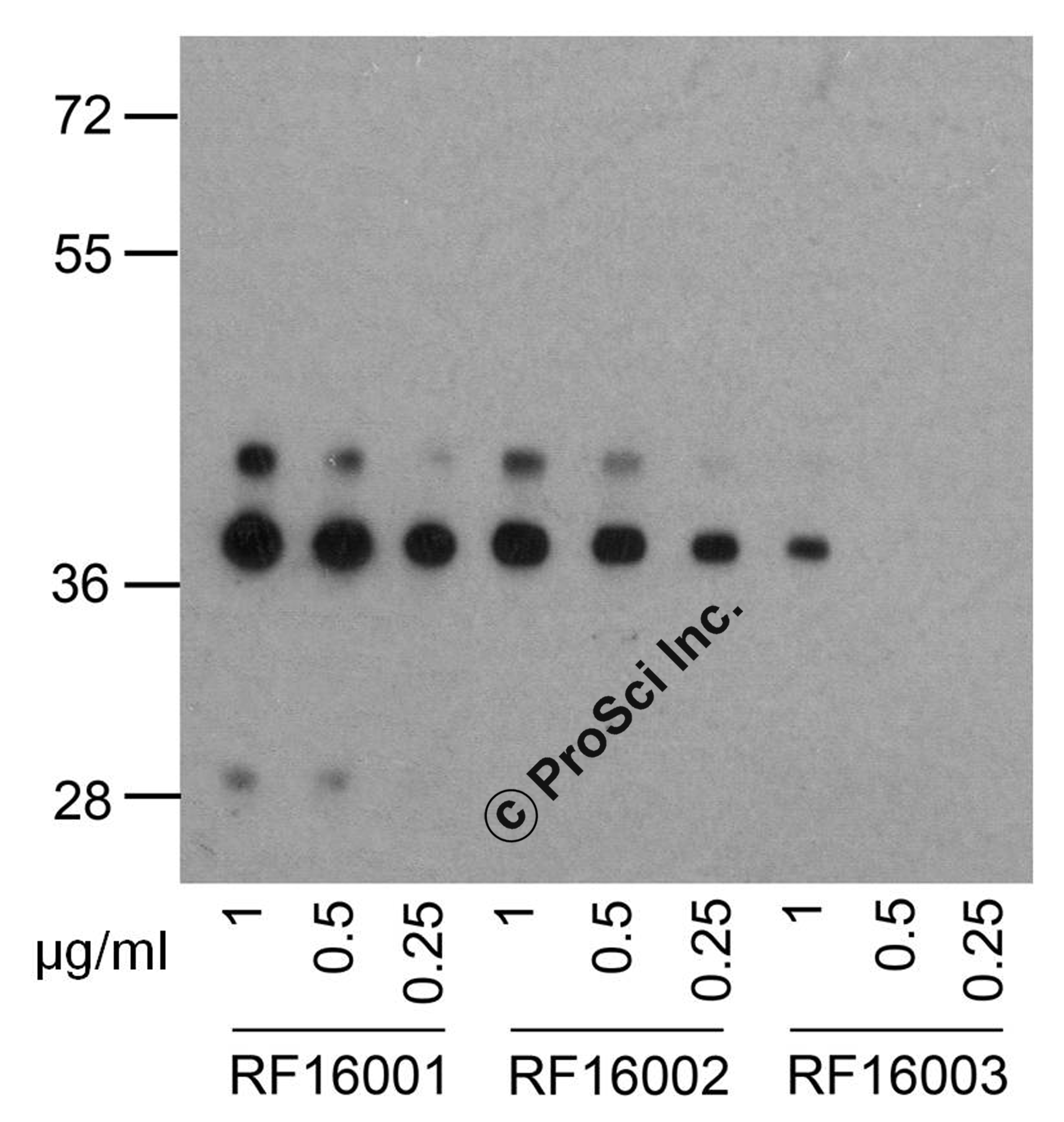
Western blot analysis of PD-1 in overexpressing 293 cells using RF16001, RF16002, and RF16003 antibody at 1, 0.5, and 0.25 μg/ml, respectively.
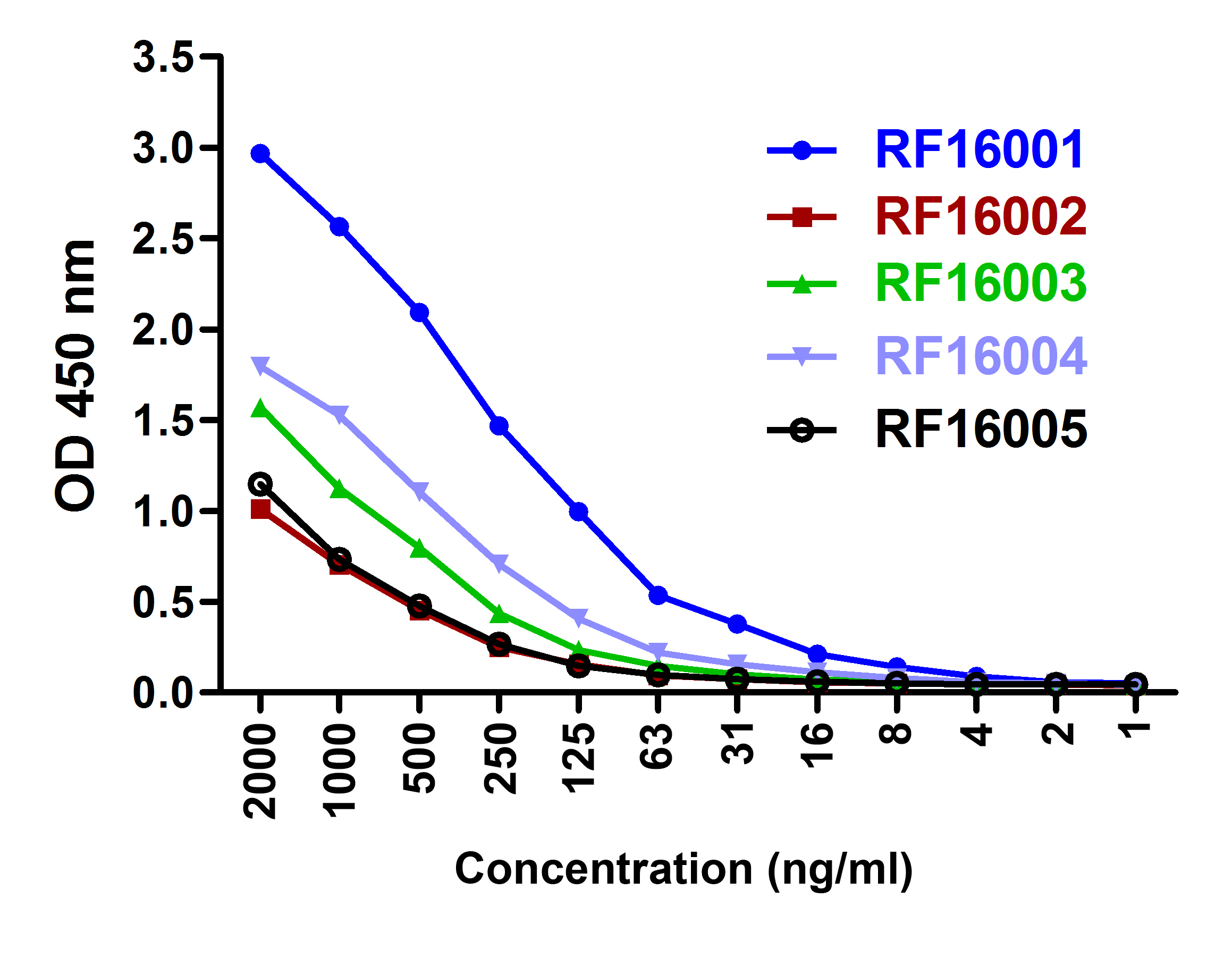
Titration curve analysis of PD-1 mAbs to detect recombinant PD-1 in ELISA with RF16001, RF16002, RF16003, RF16004, and RF16005 abs at decreasing concentrations.
|
|
|
|
Western blot analysis of PD-1 in transfected 293 cell lysate with PD-1 antibody at (A) 0.5 and (B) 1 μg/mL.
|
|
| 別品名 |
PD-1 Antibody: PD1, PD-1, CD279, SLEB2, hPD-1, hPD-l, hSLE1, PD1, Programmed cell death protein 1, Protein PD-1, PDCD1, PDCD-1
|
| 交差種 |
Human
|
| 適用 |
Western Blot
IHC paraffin embedding section
Enzyme Linked Immunosorbent Assay
Immuno Fluorescence
Immunocytochemistry (cell)
|
| 免疫動物 |
Mouse
|
| クローン |
8A4
|
| 抗体クラス |
IgG1
|
| 標識物 |
Unlabeled
|
| 精製度 |
Ig fraction - Protein A
|
| GENE ID |
5133
|
| Accession No.(Gene/Protein) |
NP_005009
|
| Gene Symbol |
PDCD1
|
| その他 |
[Protein GI Number]145559515
[Swiss-Prot No]Q15116
|
| 参考文献 |
Holling TM, Schooten E, and van Den Elsing PJ. Function and regulation of MHC class II molecules in T-lymphocytes: of mice and men. Hum. Immunol. 2004; 65:282-90.
Ishida Y, Agata Y, Shibahara K, et al. Induced expression of PD-1, a novel member of the immunoglobulin gene superfamily, upon programmed cell death. EMBO J. 1992; 11:3887-95.
Zhong X, Bai C, Gao W, et al. Suppression of expression and function of negative immune regulator PD-1 by certain pattern recognition and cytokine receptor signals associated with immune system danger. Int. Immunol. 2004; 16:1181-8.
Nishimura H, Nose M, Hiai H, et al. Development of lupus-like autoimmune diseases by the disruption of the PD-1 gene encoding an ITIM motif-carrying immunoreceptor. Immunity 1999; 11:141-51.
|
|
| メーカー |
品番 |
包装 |
|
PSC
|
RF16002
|
0.1 MG
[1 mg/mL]
|
※表示価格について
| 当社在庫 |
なし
|
| 納期目安 |
3週間程度
|
| 保存温度 |
-20℃
|
|
※当社では商品情報の適切な管理に努めておりますが、表示される法規制情報は最新でない可能性があります。
また法規制情報の表示が無いものは、必ずしも法規制に非該当であることを示すものではありません。
商品のお届け前に最新の製品法規制情報をお求めの際はこちらへお問い合わせください。
|
※当社取り扱いの試薬・機器製品および受託サービス・創薬支援サービス(納品物、解析データ等)は、研究用としてのみ販売しております。
人や動物の医療用・臨床診断用・食品用としては、使用しないように、十分ご注意ください。
法規制欄に体外診断用医薬品と記載のものは除きます。
|
|
※リンク先での文献等のダウンロードに際しましては、掲載元の規約遵守をお願いします。
|
|
※CAS Registry Numbers have not been verified by CAS and may be inaccurate.
|
