|
※サムネイル画像をクリックすると拡大画像が表示されます。
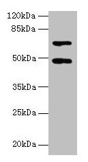
Western blot
All lanes: HDAC3 antibody at 2μg/ml + Hela whole cell lysate
Secondary
Goat polyclonal to rabbit IgG at 1/10000 dilution
Predicted band size: 49, 50 kDa
Observed band size: 49 kDa
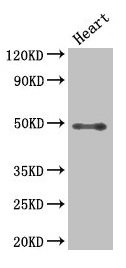
Western Blot
Positive WB detected in: Mouse heart tissue
All lanes: HDAC3 antibody at 2.5μg/ml
Secondary
Goat polyclonal to rabbit IgG at 1/50000 dilution
Predicted band size: 49, 50 kDa
Observed band size: 49 kDa
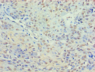
Immunohistochemistry of paraffin-embedded human breast cancer using CSB-PA010239LA01HU at dilution of 1:100
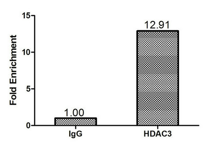
Chromatin Immunoprecipitation Hela(1.2*106)were cross-linked with formaldehyde, sonicated, and immunoprecipitated with 4μg anti-HDAC3 or a control normal rabbit IgG. The resulting ChIP DNA was quantified tissue using real-time PCR with primers(CSB-PP010239HU) against the P21 promoter.
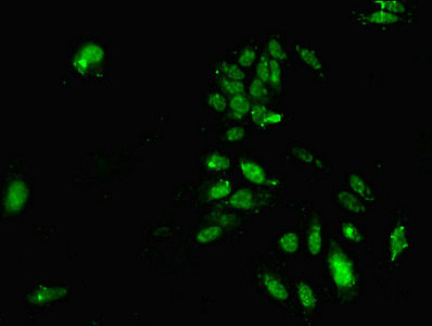
Immunofluorescent analysis of Hela cells using CSB-PA010239LA01HU at dilution of 1:100 and Alexa Fluor 488-congugated AffiniPure Goat Anti-Rabbit IgG(H+L)
|
|
|
|
Western blot
All lanes: HDAC3 antibody at 2μg/ml + Hela whole cell lysate
Secondary
Goat polyclonal to rabbit IgG at 1/10000 dilution
Predicted band size: 49, 50 kDa
Observed band size: 49 kDa
|
|
| 別品名 |
HD3 antibody; HDAC 3 antibody; HDAC3 antibody; HDAC3_HUMAN antibody; Histone deacetylase 3 antibody; RPD3 2 antibody; RPD3 antibody; RPD3-2 antibody; SMAP45 antibody
|
| 種由来 |
Human
|
| 交差種 |
Human
Mouse
|
| 適用 |
Western Blot
Enzyme Linked Immunosorbent Assay
Immunohistochemistry
Immuno Fluorescence
Chromatin Immunoprecipitation
|
| 免疫動物 |
Rabbit
|
| 抗体クラス |
IgG
|
| 抗原部位 |
a.a.1-428
|
| 標識物 |
Unlabeled
|
| 精製度 |
Ig fraction - Protein G
|
| Accession No.(Gene/Protein) |
O15379
|
| 参考文献 |
[1]"A hybrid mechanism of action for BCL6 in B cells defined by formation of functionally distinct complexes at enhancers and promoters." Hatzi K., Jiang Y., Huang C., Garrett-Bakelman F., Gearhart M.D., Giannopoulou E.G., Zumbo P., Kirouac K., Bhaskara S., Polo J.M., Kormaksson M., Mackerell A.D. Jr., Xue F., Mason C.E., Hiebert S.W., Prive G.G., Cerchietti L., Bardwell V.J., Elemento O., Melnick A. Cell Rep. 4:578-588(2013) . [2]"Acetylation of a conserved lysine residue in the ATP binding pocket of p38 augments its kinase activity during hypertrophy of cardiomyocytes." Pillai V.B., Sundaresan N.R., Samant S.A., Wolfgeher D., Trivedi C.M., Gupta M.P. Mol. Cell. Biol. 31:2349-2363(2011). [3]"Initial characterization of the human central proteome." Burkard T.R., Planyavsky M., Kaupe I., Breitwieser F.P., Buerckstuemmer T., Bennett K.L., Superti-Furga G., Colinge J. BMC Syst. Biol. 5:17-17(2011).
|
|
| メーカー |
品番 |
包装 |
|
CSB
|
CSB-PA010239LA01HU
|
50 UG
|
※表示価格について
| 当社在庫 |
なし
|
| 納期目安 |
2週間程度
|
| 保存温度 |
-20℃
|
|
※当社では商品情報の適切な管理に努めておりますが、表示される法規制情報は最新でない可能性があります。
また法規制情報の表示が無いものは、必ずしも法規制に非該当であることを示すものではありません。
商品のお届け前に最新の製品法規制情報をお求めの際はこちらへお問い合わせください。
|
※当社取り扱いの試薬・機器製品および受託サービス・創薬支援サービス(納品物、解析データ等)は、研究用としてのみ販売しております。
人や動物の医療用・臨床診断用・食品用としては、使用しないように、十分ご注意ください。
法規制欄に体外診断用医薬品と記載のものは除きます。
|
|
※リンク先での文献等のダウンロードに際しましては、掲載元の規約遵守をお願いします。
|
|
※CAS Registry Numbers have not been verified by CAS and may be inaccurate.
|
