
| 別品名 |
Eukaryotic translation elongation factor 1 alpha 1, CCS-3, CCS3, EEF-1, EEF1A, eEF1A-1, EF-Tu, EF1A, FLJ25721, GRAF-1EF, HNGC:16303, PTI1
|
| 抗原部位 |
a.a.1-462
|
| 種由来 |
Human
|
| 標識物 |
Unlabeled
|
| 精製度 |
Ig fraction - Protein A
|
| 適用 |
Western Blot
Enzyme Linked Immunosorbent Assay
Immuno Fluorescence
Immunocytochemistry (cell)
|
| 免疫動物 |
Mouse
|
| 抗体クラス |
IgG2aκ
|
| クローン |
AT23C11
|
| 交差種 |
Human
|
| Accession No.(Gene/Protein) |
NP_001393, P68104
|
| 形状 |
液状
|
| 参考文献 |
Huang Y., et al. (2012) Zhonqquo Shi Yan Xue Ye Xue Za Zhi. 20(4): 835-841.
Browne GJ., et al. (2002) Eur J Biochem. 269(22): 5360-5368.
|
|
※サムネイル画像をクリックすると拡大画像が表示されます。
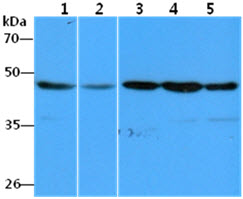
The Cell lysates (40ug) were resolved by SDS-PAGE, transferred to PVDF membrane and probed with anti-human EEF1A1 antibody (1:1000). Proteins were visualized using a goat anti-mouse secondary antibody conjugated to HRP and an ECL detection system.
Lane 1.: HeLa cell lysate
Lane 2.: A549 cell lysate
Lane 3.: Raji cell lysate
Lane 4.: THP-1 cell lysate
Lane 5.: MCF-7 cell lysate
ICC/IF analysis of EEF1A1 in A549 cells line, stained with DAPI (Blue) for nucleus staining and monoclonal anti-human EEF1A1 antibody (1:100) with goat anti-mouse IgG-Alexa fluor 488 conjugate (Green).

|
|
|
|
The Cell lysates (40ug) were resolved by SDS-PAGE, transferred to PVDF membrane and probed with anti-human EEF1A1 antibody (1:1000). Proteins were visualized using a goat anti-mouse secondary antibody conjugated to HRP and an ECL detection system.
Lane 1.: HeLa cell lysate
Lane 2.: A549 cell lysate
Lane 3.: Raji cell lysate
Lane 4.: THP-1 cell lysate
Lane 5.: MCF-7 cell lysate
|
|
|
| メーカー |
品番 |
包装 |
|
ATG
|
ATGA0347
|
100 UL
[1mg/ml]
|
※表示価格について
| 当社在庫 |
なし
|
| 納期目安 |
1週間程度
|
| 保存温度 |
-70℃
|
|

