Fig.1 Western blot analysis
Whole cell lysaes prepared from DJM-1 cells (lane 1) and HeLa cells (lane 2) were
immunoblotted with PN753 or PC742 at 1:200 dilution.
Plectin antibodies detected approximate 500 kDa bands in these cell lysates (arrow). Smaller polypeptide found in lane 2 may be a degraded product or alternatively spliced rod-less isoform of plectin. Polypeptiedes were separated by SDS-PAGE (5% separating gel).
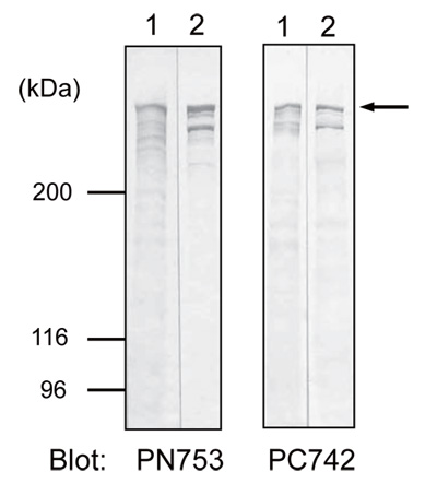
Fig.2 Location of the epitopes for the plectin antibodies
PN753 and PC742 clones were obtained by immunizing
mice with the NH2- (173-595aa) or the COOH-terminal
(2,930-3,153aa) regions of human plectin (4,574aa),
respectively. Gray box represents a predicted coiled-coil
region (1,300-2,600aa).
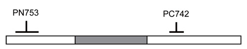
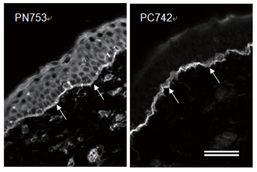
Fig.3 Immunofluorescence microscopy of human skin
Human skin sections were stained with PC742 (1:100
dilution) or PN753 (1:100 dilution). Arrows indicate
dermal-epidermal junctions. PN753 stains epidermal cells
in addition to hemidesmosomes at the dermal-epidermal
junction. Sections were fixed with .20℃ acetone for 10
min. Bar: 50um.