Anti BP180/type XVII collagen
|


| Һн—R—Ҳ |
Human
|
| “K—p |
Western Blot
Immuno Fluorescence
Immunoprecipitation
|
| –Жүu“®•Ё |
Mouse
|
| ғNғҚҒ[ғ“ |
C34
|
| ҢрҚ·Һн |
Human
|
|
ҒҰғTғҖғlғCғӢүж‘ңӮрғNғҠғbғNӮ·ӮйӮЖҠg‘еүж‘ңӮӘ•\ҺҰӮіӮкӮЬӮ·ҒB
Fig.1 Western blot analysis
TritonX-100 insoluble cytoskeletal fraction (lane 1) and concentrated conditioned
medium (lane 2) prepared from DJM-1 cells were immunoblotted with the C34 antibody (1:200 dilution).
The C34 antibody detected a band at approximately 180 kDa in lane 1. This antibody also reacted with a 120-kDa shed ectodomain of BP180 in lane 2. Polypeptiedes were separated by SDS-PAGE (7.5% separating gel).
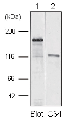
Fig.3 Immunofluorescence microscopy of human skin
A human skin section was stained with C34 antibody at 1:200 dilution. The antibody revealed the location of BP180 molecules at the dermal-epidermal junction (arrow). E: epidermis, D: dermis. Bar = 50 um. Frozen sections were prepared as described
previously (ref. 3).
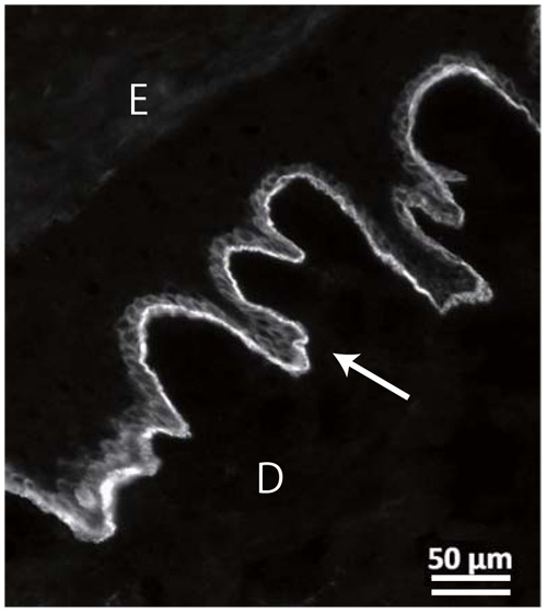
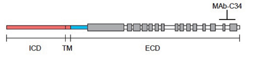
Fig.2 Location of the epitope for the C34 antibody
The C34 antibody does not react with the 100-kDa extracellular
fragment of BP180, which lacks the COOH-terminal portion
(ref. 1). The result indicates that the C34 antibody recognizes an
epitope locates at the COOH-terminal portion of about 20 kDa.
ICD, TM and ECD represent for intracellular, transmembrane
and extracellular domains. Collagenous domains are shown by
gray boxes.
|
|
|
|
Fig.1 Western blot analysis
TritonX-100 insoluble cytoskeletal fraction (lane 1) and concentrated conditioned
medium (lane 2) prepared from DJM-1 cells were immunoblotted with the C34 antibody (1:200 dilution).
The C34 antibody detected a band at approximately 180 kDa in lane 1. This antibody also reacted with a 120-kDa shed ectodomain of BP180 in lane 2. Polypeptiedes were separated by SDS-PAGE (7.5% separating gel).
|
|
|
| ғҒҒ[ғJҒ[ |
•i”Ф |
•п‘• |
|
CAC
|
NU-01-BP2
|
500 UL
|
ҒҰ•\ҺҰүҝҠiӮЙӮВӮўӮД
| “–ҺРҚЭҢЙ |
Ӯ Ӯи
|
| •Ы‘¶ү·“x |
-ӮQӮOҒҺ
|
| ғҒҒ[ғJҒ[Ҹо•с |
| ғҒҒ[ғJҒ[–ј |
ғRғXғӮҒEғoғCғIҠ”Һ®үпҺР
|
| —ӘҚҶ |
CAC
|
|
ҒҰ“–ҺРӮЕӮНҸӨ•iҸо•сӮМ“KҗШӮИҠЗ—қӮЙ“wӮЯӮДӮЁӮиӮЬӮ·ӮӘҒA•\ҺҰӮіӮкӮй–@ӢKҗ§Ҹо•сӮНҚЕҗVӮЕӮИӮўүВ”\җ«ӮӘӮ ӮиӮЬӮ·ҒB
Ғ@ӮЬӮҪ–@ӢKҗ§Ҹо•сӮМ•\ҺҰӮӘ–іӮўӮаӮМӮНҒA•KӮёӮөӮа–@ӢKҗ§ӮЙ”сҠY“–ӮЕӮ ӮйӮұӮЖӮрҺҰӮ·ӮаӮМӮЕӮНӮ ӮиӮЬӮ№ӮсҒB
Ғ@ҸӨ•iӮМӮЁ“НӮҜ‘OӮЙҚЕҗVӮМҗ»•i–@ӢKҗ§Ҹо•сӮрӮЁӢҒӮЯӮМҚЫӮНӮұӮҝӮзӮЦӮЁ–вӮўҚҮӮнӮ№ӮӯӮҫӮіӮўҒB
Ғ@
|
ҒҰ“–ҺРҺжӮиҲөӮўӮМҺҺ–тҒEӢ@Ҡнҗ»•iӮЁӮжӮСҺу‘хғTҒ[ғrғXҒE‘n–тҺxүҮғTҒ[ғrғXҒi”[•i•ЁҒAүрҗНғfҒ[ғ^“ҷҒjӮНҒAҢӨӢҶ—pӮЖӮөӮДӮМӮЭ”М”„ӮөӮДӮЁӮиӮЬӮ·ҒB
Ғ@җlӮв“®•ЁӮМҲг—Г—pҒE—ХҸ°җf’f—pҒEҗH•i—pӮЖӮөӮДӮНҒAҺg—pӮөӮИӮўӮжӮӨӮЙҒAҸ\•ӘӮІ’ҚҲУӮӯӮҫӮіӮўҒB
Ғ@–@ӢKҗ§—“ӮЙ‘МҠOҗf’f—pҲг–т•iӮЖӢLҚЪӮМӮаӮМӮНҸңӮ«ӮЬӮ·ҒB
|
|
ҒҰғҠғ“ғNҗжӮЕӮМ•¶ҢЈ“ҷӮМғ_ғEғ“ғҚҒ[ғhӮЙҚЫӮөӮЬӮөӮДӮНҒAҢfҚЪҢіӮМӢK–сҸ…ҺзӮрӮЁҠиӮўӮөӮЬӮ·ҒB
|

