|
※サムネイル画像をクリックすると拡大画像が表示されます。
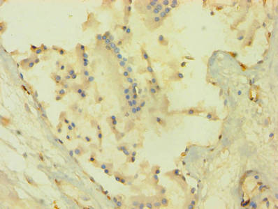
Immunohistochemistry of paraffin-embedded human prostate tissue using CSB-PA15905A0Rb at dilution of 1:100
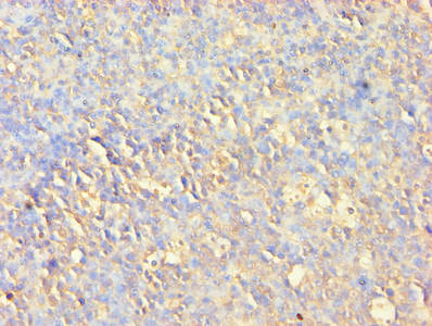
Immunohistochemistry of paraffin-embedded human tonsil tissue using CSB-PA15905A0Rb at dilution of 1:100
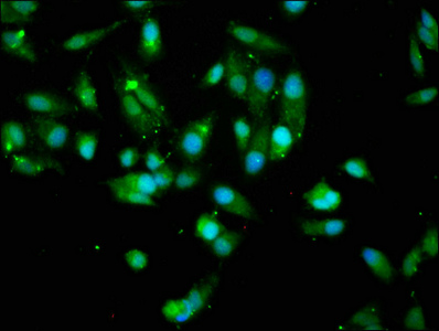
Immunofluorescence staining of Hela cells with CSB-PA15905A0Rb at 1:200, counter-stained with DAPI. The cells were fixed in 4% formaldehyde, permeabilized using 0.2% Triton X-100 and blocked in 10% normal Goat Serum. The cells were then incubated with the antibody overnight at 4°C. The secondary antibody was Alexa Fluor 488-congugated AffiniPure Goat Anti-Rabbit IgG(H+L).
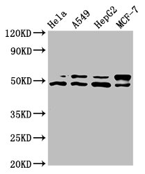
Western Blot
Positive WB detected in: Hela whole cell lysate, A549 whole cell lysate, HepG2 whole cell lysate, MCF-7 whole cell lysate
All lanes: AKT1 antibody at 7.4μg/ml
Secondary
Goat polyclonal to rabbit IgG at 1/50000 dilution
Predicted band size: 56, 49 kDa
Observed band size: 56, 49 kDa
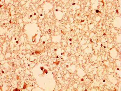
IHC image of CSB-PA15905A0Rb diluted at 1:200 and staining in paraffin-embedded human brain tissue performed on a Leica BondTM system. After dewaxing and hydration, antigen retrieval was mediated by high pressure in a citrate buffer (pH 6.0). Section was blocked with 10% normal goat serum 30min at RT. Then primary antibody (1% BSA) was incubated at 4°C overnight. The primary is detected by a biotinylated secondary antibody and visualized using an HRP conjugated SP system.
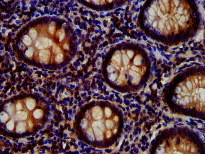
IHC image of CSB-PA15905A0Rb diluted at 1:700 and staining in paraffin-embedded human appendix tissue performed on a Leica BondTM system. After dewaxing and hydration, antigen retrieval was mediated by high pressure in a citrate buffer (pH 6.0). Section was blocked with 10% normal goat serum 30min at RT. Then primary antibody (1% BSA) was incubated at 4°C overnight. The primary is detected by a biotinylated secondary antibody and visualized using an HRP conjugated SP system.
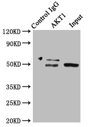
Immunoprecipitating AKT1 in HepG2 whole cell lysate
Lane 1: Rabbit control IgG instead of CSB-PA15905A0Rb in HepG2 whole cell lysate. For western blotting, a HRP-conjugated Protein G antibody was used as the secondary antibody (1/2000)
Lane 2: CSB-PA15905A0Rb (8μg) + HepG2 whole cell lysate (500μg)
Lane 3: HepG2 whole cell lysate (20μg)
|
|
|
|
Immunohistochemistry of paraffin-embedded human prostate tissue using CSB-PA15905A0Rb at dilution of 1:100
|
|
| 別品名 |
AKT 1 antibody; AKT antibody; AKT1 antibody; AKT1_HUMAN antibody; C AKT antibody; cAKT antibody; MGC99656 antibody; PKB alpha antibody; PKB antibody; PKB-ALPHA antibody; PRKBA antibody; Protein Kinase B Alpha antibody; Protein kinase B antibody; Proto-oncogene c-Akt antibody; RAC Alpha antibody; RAC antibody; Rac protein kinase alpha antibody; RAC Serine/Threonine Protein Kinase antibody; RAC-alpha serine/threonine-protein kinase antibody; RAC-PK-alpha antibody; v akt murine thymoma viral oncogene homolog 1 antibody; vAKT Murine Thymoma Viral Oncogene Homolog 1 antibody
|
| 種由来 |
Human
|
| 交差種 |
Human
|
| 適用 |
Western Blot
Enzyme Linked Immunosorbent Assay
Immunohistochemistry
Immuno Fluorescence
Immunoprecipitation
|
| 免疫動物 |
Rabbit
|
| 抗体クラス |
IgG
|
| 抗原部位 |
a.a.1-480
|
| 標識物 |
Unlabeled
|
| 精製度 |
Ig fraction - Protein G
|
| Accession No.(Gene/Protein) |
P31749
|
| 参考文献 |
[Pub Med ID]30288055
|
|
| メーカー |
品番 |
包装 |
|
CSB
|
CSB-PA15905A0RB
|
100 UG
|
※表示価格について
| 当社在庫 |
なし
|
| 納期目安 |
2週間程度
|
| 保存温度 |
-20℃
|
|
※当社では商品情報の適切な管理に努めておりますが、表示される法規制情報は最新でない可能性があります。
また法規制情報の表示が無いものは、必ずしも法規制に非該当であることを示すものではありません。
商品のお届け前に最新の製品法規制情報をお求めの際はこちらへお問い合わせください。
|
※当社取り扱いの試薬・機器製品および受託サービス・創薬支援サービス(納品物、解析データ等)は、研究用としてのみ販売しております。
人や動物の医療用・臨床診断用・食品用としては、使用しないように、十分ご注意ください。
法規制欄に体外診断用医薬品と記載のものは除きます。
|
|
※リンク先での文献等のダウンロードに際しましては、掲載元の規約遵守をお願いします。
|
|
※CAS Registry Numbers have not been verified by CAS and may be inaccurate.
|
