|
※サムネイル画像をクリックすると拡大画像が表示されます。
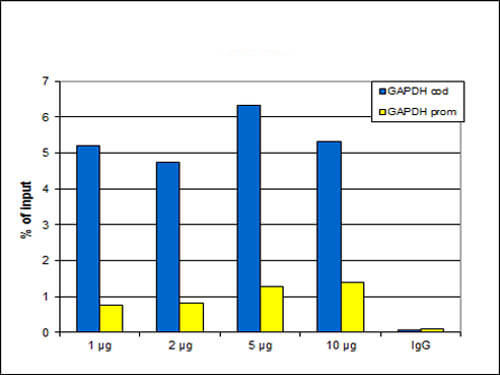
Chromatin Immunoprecipitation Rabbit Anti-H3K27me1 Antibody. ChIP assays were performed using HeLa cells, H3K27me1 Antibody, and optimized PCR primer sets for qPCR. ChIP was performed using sheared chromatin from 100,000 cells. A titration of the antibody consisting of 1, 2, 5 and 10 μg per ChIP experiment was analyzed. IgG (2 μg/IP) was used as negative IP control. qPCR was performed with primers for the promoter and the coding region of the active gene GAPDH used as a negative and a positive control target, respectively. This figure shows the recovery, expressed as a % of input (the relative amount of immunoprecipitated DNA compared to input DNA after qPCR analysis).
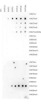
Dot Blot results of H3K27me1 antibody. Antigens: H3K27me1 and other modifications and unmodified sequences of histone H3 and H4. Load: 100 pmol, 50 pmol, 25 pmol, 5 pmol, 2 pmol, and 0.2 pmol with control. Primary antibody: H3K27me1 antibody at 1:20,000 for 45 min at 4°C. Secondary antibody: anti-rabbit HRP antibody at 1:20,000 for 45 min at RT. Block: 5% BLOTTO.
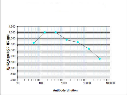
ELISA results of H3K27me1 antibody. Antigen: BSA coupled to H3K27me1. Coating amount: 0.1 μg per well. Dilution series: serial. Titer: 1:32,900 H3K27me1 antibody. Substrate: TMB (p/n TMBE-1000).
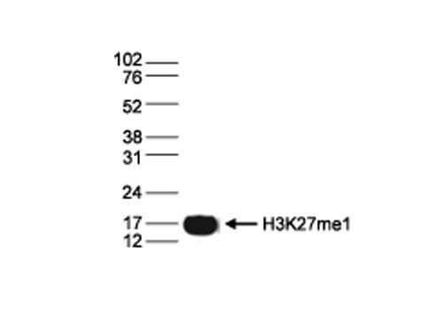
Western Blot results of Rabbit anti-Histone H3K27me1 antibody. Lane 1: 15 μg of histone extracts from HeLa cells. Primary antibody: Rabbit anti-Histone H3 K27 me2 antibody at 1:1,000. Secondary antibody: Peroxidase anti-rabbit secondary antibody at 1:10,000 for 45 min at RT. Block: TBS-Tween / 5% BLOTTO. Predicted/Observed size: ~15 kDa for Rabbit Histone H3 K27 me2.
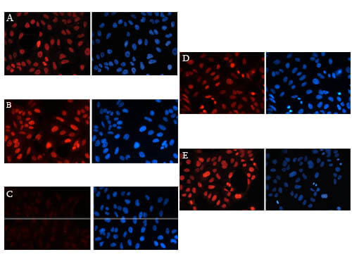
Immunofluorescence results of Anti-Histone H3K27me1 Antibody. Tissue: Human osteosarcoma (U2OS) cells. Fixation: 4% formaldehyde for 20 min. Block: PBS/Triton X-100 / 5% normal goat serum. Incubation: Figure A-E: H3K27me1 antibody 1:1,000. Figure B: 2ng/μl H3K27 peptide. Figure C: 2ng/μl H3K27me1 peptide. Figure D: 2ng/μl H3K27me2 peptide. Figure E: 2ng/μl H3K27me3 peptide. Secondary antibody: anti-rabbit Alexa568 secondary antibody at 1:10,000 for 45 min at RT (Left) or DAPI (right). Staining: Histone H3K27me1 antibody as (red) fluorescent signal, DAPI (blue).
|
|
|
|
Chromatin Immunoprecipitation Rabbit Anti-H3K27me1 Antibody. ChIP assays were performed using HeLa cells, H3K27me1 Antibody, and optimized PCR primer sets for qPCR. ChIP was performed using sheared chromatin from 100,000 cells. A titration of the antibody consisting of 1, 2, 5 and 10 μg per ChIP experiment was analyzed. IgG (2 μg/IP) was used as negative IP control. qPCR was performed with primers for the promoter and the coding region of the active gene GAPDH used as a negative and a positive control target, respectively. This figure shows the recovery, expressed as a % of input (the relative amount of immunoprecipitated DNA compared to input DNA after qPCR analysis).
|
|
| 別品名 |
Histone H3.1, Histone H3/a, Histone H3/b, Histone H3/c, Histone H3/d, Histone H3/e, Histone H3/f, Histone H3/g, Histone H3/h, Histone H3/i, Histone H3/j, Histone H3/k, Histone H3/l
|
| 交差種 |
Human
|
| 適用 |
Western Blot
Enzyme Linked Immunosorbent Assay
Immuno Fluorescence
Chromatin Immunoprecipitation
Dot Blot
|
| 免疫動物 |
Rabbit
|
| 抗体クラス |
IgG
|
| 標識物 |
Unlabeled
|
| 精製度 |
Affinity Purified
|
| GENE ID |
3020
|
| Accession No.(Gene/Protein) |
NP_002098, P84243
|
| Gene Symbol |
HIST1H3A
|
| [注意事項] |
濃度はロットによって異なる可能性があります。メーカーDS及びCoAからご確認ください。
|
|
| メーカー |
品番 |
包装 |
|
RKL
|
600-401-W54
|
50 UG
|
※表示価格について
| 当社在庫 |
なし
|
| 納期目安 |
約10日
|
| 保存温度 |
-20℃
|
|
※当社では商品情報の適切な管理に努めておりますが、表示される法規制情報は最新でない可能性があります。
また法規制情報の表示が無いものは、必ずしも法規制に非該当であることを示すものではありません。
商品のお届け前に最新の製品法規制情報をお求めの際はこちらへお問い合わせください。
|
※当社取り扱いの試薬・機器製品および受託サービス・創薬支援サービス(納品物、解析データ等)は、研究用としてのみ販売しております。
人や動物の医療用・臨床診断用・食品用としては、使用しないように、十分ご注意ください。
法規制欄に体外診断用医薬品と記載のものは除きます。
|
|
※リンク先での文献等のダウンロードに際しましては、掲載元の規約遵守をお願いします。
|
