|
※サムネイル画像をクリックすると拡大画像が表示されます。
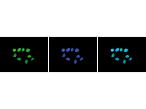
Immunofluorescence Microscopy results of Rabbit anti-Histone H3 K27 me2 antibody. Tissue: HeLa cells. Fixation: 4% formaldehyde for 10 min. Block: PBS/Triton X-100 / 5% normal goat serum and 1% BSA. Primary antibody: Histone H3 K27 me2 antibody at 1:500 for 1 hr at RT. Secondary antibody: anti-rabbit Alexa 488 secondary antibody at 1:10,000 for 45 min at RT. Staining: Histone H3 K27 me2 antibody as green fluorescent signal (left), with DAPI (middle), and a merge of the two stainings (right).
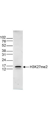
Western Blot results of Rabbit anti-Histone H3 K27 me2 antibody. Lane 1: 15 μg of histone extracts from HeLa cells. Primary antibody: Rabbit anti-Histone H3 K27 me2 antibody at 1:1,000 overnight at 4°C. Secondary antibody: Peroxidase anti-rabbit secondary antibody at 1:10,000 for 45 min at RT. Block: TBS-Tween / 5% BLOTTO. Predicted/Observed size: ~15 kDa for Rabbit Histone H3 K27 me2.
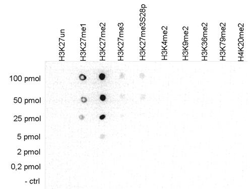
Dot Blot results of Rabbit anti-Histone H3 K27 me2 antibody. Antigens: H3K27, H3K27me1, H3K27me2, H3K27me3, H3K27me3 S28p, H3K4me2, H3K9me2, H3K36me2, H3K79me2, H4K20me2. Load: 100pmol, 50pmol, 25pmol, 5pmol, 2pmol, 0.2pmol, and control. Primary antibody: Histone H3 K27 me2 antibody at 1:50,000 for 45 min at 4°C. Secondary antibody: anti-rabbit HRP secondary antibody at 1:10,000 for 45 min at RT. Block: 5% BLOTTO.
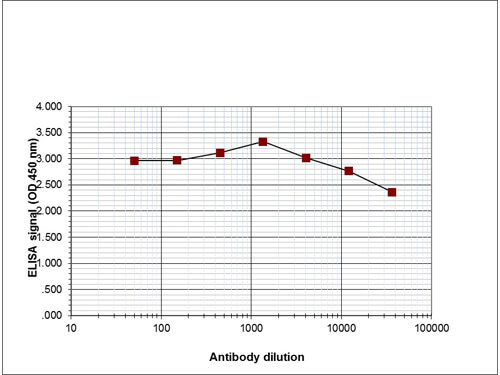
ELISA results of Rabbit anti-Histone H3 K27 me2 antibody. Antigen: Histone H3 K27 me2. Coating amount: 0.1 μg per well. Dilution series: serial. Titer: 1:480,000 Histone H3 K27 me2 antibody. Substrate: TMB (p/n TMBE-1000).
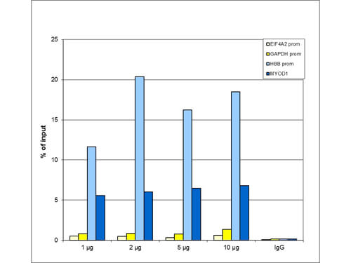
Chromatin Immunoprecipitation Rabbit H3K27me2 Antibody. ChIP assays were performed using HeLa cells, the Anti-H3K27me2 Antibody, and optimized PCR primer sets for qPCR. ChIP was performed using sheared chromatin from 1 million cells. A titration of the antibody consisting of 1, 2, 5, and 10 μg per ChIP experiment was analyzed. IgG (2 μg/IP) was used as negative IP control. QPCR was performed with primers for the promoter of the active GAPDH and EIF4A2 genes, used as negative controls, and for the promoter of the inactive HBB and the coding region of the inactive MYOD1 genes, used as positive controls. This figure shows the recovery, expressed as a % of input (the relative amount of immunoprecipitated DNA compared to input DNA after qPCR analysis). These results are in accordance with the observation that H3K27me2 is preferably present at silent genes.
|
|
|
|
Immunofluorescence Microscopy results of Rabbit anti-Histone H3 K27 me2 antibody. Tissue: HeLa cells. Fixation: 4% formaldehyde for 10 min. Block: PBS/Triton X-100 / 5% normal goat serum and 1% BSA. Primary antibody: Histone H3 K27 me2 antibody at 1:500 for 1 hr at RT. Secondary antibody: anti-rabbit Alexa 488 secondary antibody at 1:10,000 for 45 min at RT. Staining: Histone H3 K27 me2 antibody as green fluorescent signal (left), with DAPI (middle), and a merge of the two stainings (right).
|
|
| 別品名 |
Histone H3.1, Histone H3/a, Histone H3/b, Histone H3/c, Histone H3/d, Histone H3/e, Histone H3/f, Histone H3/g, Histone H3/h, Histone H3/i, Histone H3/j, Histone H3/k, Histone H3/l
|
| 交差種 |
Human
Zebrafish
|
| 適用 |
Western Blot
Enzyme Linked Immunosorbent Assay
Immuno Fluorescence
Chromatin Immunoprecipitation
Dot Blot
|
| 免疫動物 |
Rabbit
|
| 抗体クラス |
IgG
|
| 標識物 |
Unlabeled
|
| 精製度 |
Affinity Purified
|
| GENE ID |
3020
|
| Accession No.(Gene/Protein) |
NP_002098, P84243
|
| Gene Symbol |
HIST1H3A
|
| 参考文献 |
Chaturvedi CP, Hosey AM, Palii C, Perez-Iratxeta C, Nakatani Y, Ranish JA, Dilworth FJ, and Brand M (2009) Dual role for the methyltransferase G9a in the maintenance of beta-globin gene transcription in adult erythroid cells. PNAS 106: 18303-18308.
|
| [注意事項] |
濃度はロットによって異なる可能性があります。メーカーDS及びCoAからご確認ください。
|
|
| メーカー |
品番 |
包装 |
|
RKL
|
600-401-V34
|
50 UG
|
※表示価格について
| 当社在庫 |
なし
|
| 納期目安 |
約10日
|
| 保存温度 |
-20℃
|
|
