|
※サムネイル画像をクリックすると拡大画像が表示されます。
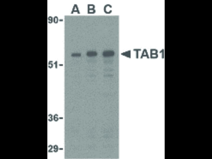
Western Blot of TAB1 antibody in 3T3 cell lysate. Lane A: TAB1 antibody at 0.5 μg/mL. Lane B: TAB1 antibody at 1 μg/mL. Lane C: TAB1 antibody at 2 μg/mL. Load: 35 μg per lane. Primary antibody: TAB1 antibody at designated concentrations for overnight at 4°C. Secondary antibody: Peroxidase rabbit secondary antibody at 1:10,000 for 45 min at RT. Block: 5% BLOTTO overnight at 4°C. Predicted/Observed size: 54 kDa, 55 kDa for TAB1. Other band(s): TAB1 splice variants and isoforms.
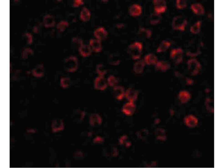
Immunofluorescence Microscopy of TAB1 antibody. Cell Type: 3T3 cells. Fixation: 0.5% PFA. Antigen retrieval: not required. Primary antibody: TAB1 antibody at 2 μg/mL for 1 h at RT. Secondary antibody: Fluorescein rabbit secondary antibody at 1:10,000 for 45 min at RT. Localization: TAB1 is located in the cell junction, cell membrane, synapse, and synaptosome. Staining: TAB1 as red fluorescent signal.
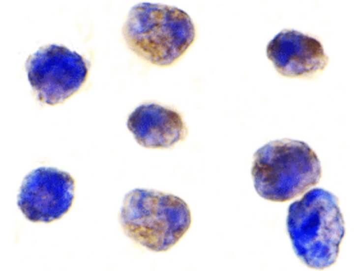
Immunocytochemistry of TAB1 antibody. Cell Type: K562 cells. Fixation: formalin fixed paraffin embedded. Antigen retrieval: not required. Primary antibody: TAB1 antibody at 1 μg/mL for 1 h at RT. Secondary antibody: Peroxidase rabbit secondary antibody at 1:10,000 for 45 min at RT. Localization: TAB1 is located in the cell junction, cell membrane, synapse, and synaptosome. Staining: TAB1 is stained brown with hematoxylin purple counterstain.
|
|
|
|
Western Blot of TAB1 antibody in 3T3 cell lysate. Lane A: TAB1 antibody at 0.5 μg/mL. Lane B: TAB1 antibody at 1 μg/mL. Lane C: TAB1 antibody at 2 μg/mL. Load: 35 μg per lane. Primary antibody: TAB1 antibody at designated concentrations for overnight at 4°C. Secondary antibody: Peroxidase rabbit secondary antibody at 1:10,000 for 45 min at RT. Block: 5% BLOTTO overnight at 4°C. Predicted/Observed size: 54 kDa, 55 kDa for TAB1. Other band(s): TAB1 splice variants and isoforms.
|
|
| 別品名 |
TGF-beta-activated kinase 1 and MAP3K7-binding protein 1, Mitogen-activated protein kinase kinase kinase 7-interacting protein 1, TGF-beta-activated kinase 1-binding protein 1, TAK1-binding protein 1, TAB1, MAP3K7IP1
|
| 交差種 |
Human
Mouse
|
| 適用 |
Western Blot
Enzyme Linked Immunosorbent Assay
Immunohistochemistry
Immuno Fluorescence
|
| 免疫動物 |
Rabbit
|
| 標識物 |
Unlabeled
|
| 精製度 |
Affinity Purified
|
| GENE ID |
10454
|
| Accession No.(Gene/Protein) |
NP_006107, Q15750
|
| Gene Symbol |
TAB1
|
| [注意事項] |
濃度はロットによって異なる可能性があります。メーカーDS及びCoAからご確認ください。
|
|
| メーカー |
品番 |
包装 |
|
RKL
|
600-401-EZ1
|
100 UG
|
※表示価格について
| 当社在庫 |
なし
|
| 納期目安 |
約10日
|
| 保存温度 |
-20℃
|
|
※当社では商品情報の適切な管理に努めておりますが、表示される法規制情報は最新でない可能性があります。
また法規制情報の表示が無いものは、必ずしも法規制に非該当であることを示すものではありません。
商品のお届け前に最新の製品法規制情報をお求めの際はこちらへお問い合わせください。
|
※当社取り扱いの試薬・機器製品および受託サービス・創薬支援サービス(納品物、解析データ等)は、研究用としてのみ販売しております。
人や動物の医療用・臨床診断用・食品用としては、使用しないように、十分ご注意ください。
法規制欄に体外診断用医薬品と記載のものは除きます。
|
|
※リンク先での文献等のダウンロードに際しましては、掲載元の規約遵守をお願いします。
|
|
※CAS Registry Numbers have not been verified by CAS and may be inaccurate.
|
