
| 別品名 |
TAB1 Antibody, 3'-Tab1, MAP3K7IP1, TGF-beta-activated kinase 1 and MAP3K7-binding protein 1, Mitogen-activated protein kinase kinase kinase 7-interacting protein 1, TAK1-binding protein 1
|
| 種由来 |
Human
|
| 標識物 |
Unlabeled
|
| 精製度 |
Affinity Purified
|
| 適用 |
Western Blot
Enzyme Linked Immunosorbent Assay
Immunohistochemistry
Immuno Fluorescence
|
| 免疫動物 |
Rabbit
|
| 交差種 |
Human
Mouse
|
| GENE ID |
10454
|
| Accession No.(Gene/Protein) |
NP_006107, Q15750
|
| Gene Symbol |
MAP3K7IP1
|
| 形状 |
滅菌済み液状品
|
| [注意事項] |
濃度はロットによって異なる可能性があります。メーカーDS及びCoAからご確認ください。
|
|
※サムネイル画像をクリックすると拡大画像が表示されます。
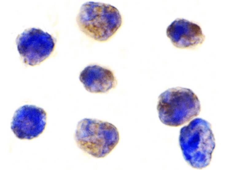
Immunocytochemistry of TAB1 antibody. Cell Type: K562 cells. Fixation: formalin fixed paraffin embedded. Antigen retrieval: not required. Primary antibody: TAB1 antibody at 1 ug/mL for 1 h at RT. Secondary antibody: Peroxidase rabbit secondary antibody at 1:10,000 for 45 min at RT. Localization: TAB1 is located in the cell junction, cell membrane, synapse, and synaptosome. Staining: TAB1 is stained brown with hematoxylin purple counterstain.
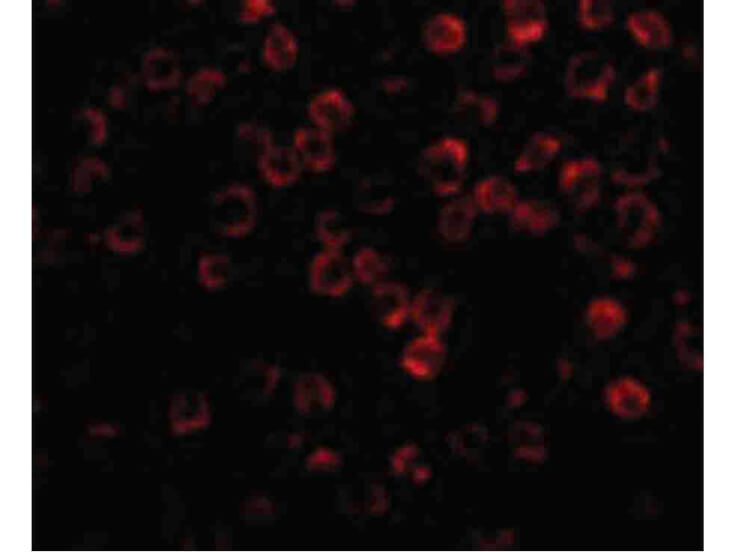
Immunofluorescence Microscopy of TAB1 antibody. Cell Type: 3T3 cells. Fixation: 0.5% PFA. Antigen retrieval: not required. Primary antibody: TAB1 antibody at 2 μg/mL for 1 h at RT. Secondary antibody: Fluorescein rabbit secondary antibody at 1:10,000 for 45 min at RT. Localization: TAB1 is located in the cell junction, cell membrane, synapse, and synaptosome. Staining: TAB1 as red fluorescent signal.
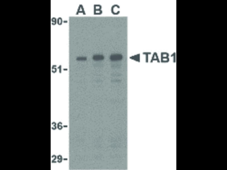
Western Blot of TAB1 antibody in 3T3 cell lysate. Lane A: TAB1 antibody at 0.5 ug/mL. Lane B: TAB1 antibody at 1 ug/mL. Lane C: TAB1 antibody at 2 ug/mL. Load: 35 ug per lane. Primary antibody: TAB1 antibody at designated concentrations for overnight at 4C. Secondary antibody: Peroxidase rabbit secondary antibody at 1:10,000 for 45 min at RT. Block: 5% BLOTTO overnight at 4C. Predicted/Observed size: 54 kDa, 55 kDa for TAB1. Other band(s): TAB1 splice variants and isoforms.
|
|
|
|
Immunocytochemistry of TAB1 antibody. Cell Type: K562 cells. Fixation: formalin fixed paraffin embedded. Antigen retrieval: not required. Primary antibody: TAB1 antibody at 1 ug/mL for 1 h at RT. Secondary antibody: Peroxidase rabbit secondary antibody at 1:10,000 for 45 min at RT. Localization: TAB1 is located in the cell junction, cell membrane, synapse, and synaptosome. Staining: TAB1 is stained brown with hematoxylin purple counterstain.
|
|
|
| メーカー |
品番 |
包装 |
|
RKL
|
600-401-EZ1
|
100 UG
|
※表示価格について
| 当社在庫 |
なし
|
| 納期目安 |
約10日
|
| 保存温度 |
-20℃
|
|

