
| 別品名 |
PDCL3 Antibody, VIAF, PHLP3, VIAF1, HTPHLP, PHLP2A, PhLP2A, Phosducin-like protein 3, VIAF-1
|
| 抗原部位 |
C-terminus
|
| 種由来 |
Human
|
| 標識物 |
Unlabeled
|
| 精製度 |
Affinity Purified
|
| 適用 |
Western Blot
Enzyme Linked Immunosorbent Assay
Immunohistochemistry
Immuno Fluorescence
|
| 免疫動物 |
Rabbit
|
| 交差種 |
Human
Mouse
Rat
|
| GENE ID |
79031
|
| Accession No.(Gene/Protein) |
NP_076970, Q9H2J4
|
| Gene Symbol |
PDCL3
|
| 形状 |
滅菌済み液状品
|
| [注意事項] |
濃度はロットによって異なる可能性があります。メーカーDS及びCoAからご確認ください。
|
|
※サムネイル画像をクリックすると拡大画像が表示されます。
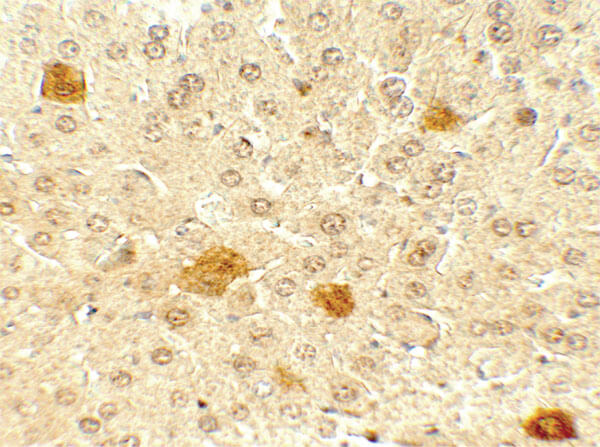
Immunohistochemistry of Rabbit anti PDCL3 antibody. Tissue: mouse liver. Primary antibody: PDCL3 antibody at 5 ug/mL. Secondary antibody: Peroxidase rabbit secondary antibody at 1:5,000. Localization: PDCL3 is secreted. Staining: PDCL3 as precipitated brown signal.
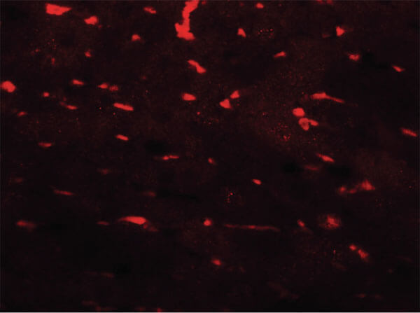
Immunofluorescence Microscopy of Rabbit anti-PDCL3 antibody. Tissue: mouse liver. Primary antibody: PDCL3 antibody at 20 μg/mL. Secondary antibody: Fluorescein rabbit secondary antibody at 1:20,000. Localization: PDCL3 is secreted. Staining: PDCL3 as red fluorescent signal.
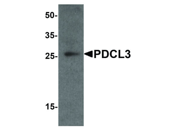
Western Blot of Rabbit anti-PDCL3 antibody. Lane A: human brain tissue lysate. Primary antibody: PDCL3 antibody at 1 ug/mL overnight at 4?C. Secondary antibody: Goat anti-Rabbit HRP secondary antibody. Block: 5% BLOTTO. Predicted/Observed size: 26 kDa, 25 kDa for PDCL3 .
|
|
|
|
Immunohistochemistry of Rabbit anti PDCL3 antibody. Tissue: mouse liver. Primary antibody: PDCL3 antibody at 5 ug/mL. Secondary antibody: Peroxidase rabbit secondary antibody at 1:5,000. Localization: PDCL3 is secreted. Staining: PDCL3 as precipitated brown signal.
|
|
|
| メーカー |
品番 |
包装 |
|
RKL
|
600-401-DK6
|
100 UG
|
※表示価格について
| 当社在庫 |
なし
|
| 納期目安 |
約10日
|
| 保存温度 |
-20℃
|
|

