
| 別品名 |
CDX2 Antibody, CDX3, CDX-3, CDX3, Homeobox protein CDX-2
|
| 抗原部位 |
C-terminus
|
| 種由来 |
Human
|
| 標識物 |
Unlabeled
|
| 精製度 |
Affinity Purified
|
| 適用 |
Western Blot
Enzyme Linked Immunosorbent Assay
Immunohistochemistry
Immuno Fluorescence
|
| 免疫動物 |
Rabbit
|
| 交差種 |
Human
Mouse
Rat
|
| GENE ID |
1045
|
| Accession No.(Gene/Protein) |
CAA74038, Q99626
|
| Gene Symbol |
CDX2
|
| 形状 |
滅菌済み液状品
|
| [注意事項] |
濃度はロットによって異なる可能性があります。メーカーDS及びCoAからご確認ください。
|
|
※サムネイル画像をクリックすると拡大画像が表示されます。
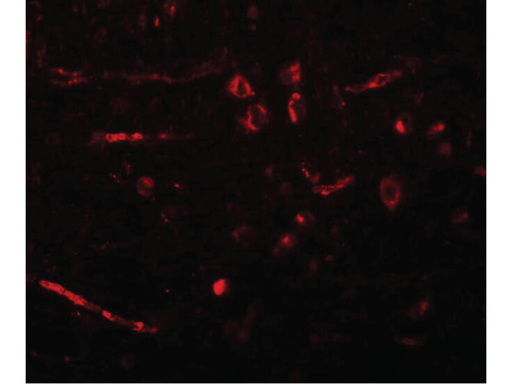
Immunofluorescence Microscopy of CDX2 antibody. Tissue: Rat brain cells. Fixation: 0.5% PFA. Antigen retrieval: not required. Primary antibody: CDX2 antibody at 20 ug/mL for 1 h at RT. Secondary antibody: Fluorescein rabbit secondary antibody at 1:10,000 for 45 min at RT. Localization: CDX2 is nuclear. Staining: CDX2 as red fluorescent signal.
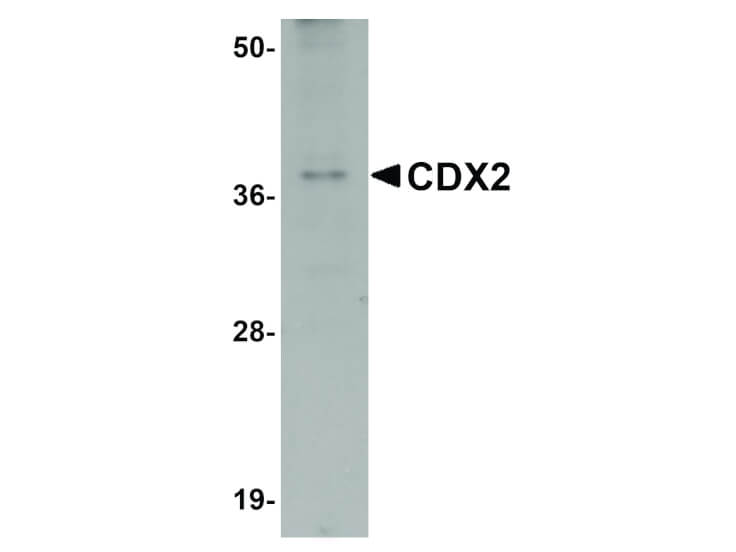
Western Blot of CDX2 antibody. Lane 1: Mouse brain tissue lysate with CDX2 antibody at 1 μg/mL. Secondary antibody: Peroxidase rabbit secondary antibody at 1:10,000 for 45 min at RT. Block: 5% BLOTTO overnight at 4°C. Predicted/Observed size: 34 kDa, 37 kDa for CDX2. Other band(s): CDX2 splice variants and isoforms.
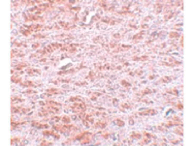
Immunohistochemistry of CDX2 antibody. Tissue: Rat brain tissue. Fixation: formalin fixed paraffin embedded. Antigen retrieval: not required. Primary antibody: CDX2 antibody at 5 ug/mL for 1 h at RT. Secondary antibody: Peroxidase rabbit secondary antibody at 1:10,000 for 45 min at RT. Localization: CDX2 is nuclear. Staining: CDX2 as precipitated pink signal with blue nuclear counterstain.
|
|
|
|
Immunofluorescence Microscopy of CDX2 antibody. Tissue: Rat brain cells. Fixation: 0.5% PFA. Antigen retrieval: not required. Primary antibody: CDX2 antibody at 20 ug/mL for 1 h at RT. Secondary antibody: Fluorescein rabbit secondary antibody at 1:10,000 for 45 min at RT. Localization: CDX2 is nuclear. Staining: CDX2 as red fluorescent signal.
|
|
|
| メーカー |
品番 |
包装 |
|
RKL
|
600-401-AH2
|
100 UG
|
※表示価格について
| 当社在庫 |
なし
|
| 納期目安 |
約10日
|
| 保存温度 |
-20℃
|
|

