

| 別品名 |
BAD Antibody, BBC2, BCL2L8, BBC6, Bcl2 antagonist of cell death, Bcl-2-binding component 6, BAD
|
| 抗原部位 |
C-terminus
|
| 種由来 |
Human
|
| 標識物 |
Unlabeled
|
| 精製度 |
Ig fraction - Ion Exchange /Gel Filtration
|
| 適用 |
Western Blot
Enzyme Linked Immunosorbent Assay
Immunohistochemistry
Immuno Fluorescence
|
| 免疫動物 |
Rabbit
|
| 交差種 |
Human
Mouse
Rat
|
| GENE ID |
572
|
| Accession No.(Gene/Protein) |
NP_004313, Q92934
|
| Gene Symbol |
BAD
|
| 形状 |
滅菌済み液状品
|
| [注意事項] |
濃度はロットによって異なる可能性があります。メーカーDS及びCoAからご確認ください。
|
|
※サムネイル画像をクリックすると拡大画像が表示されます。
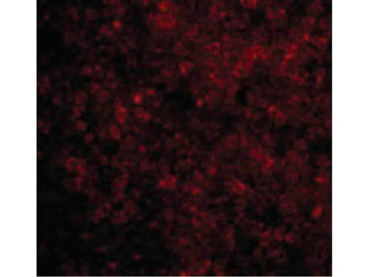
Immunofluorescence Microscopy of BAD antibody. Cell Type: rat thymus cells. Fixation: 0.5% PFA. Antigen retrieval: not required. Primary antibody: BAD antibody at 10 ug/mL for 1 h at RT. Secondary antibody: Fluorescein rabbit secondary antibody at 1:10,000 for 45 min at RT. Localization: BAD is located in the cytoplasm, cell membrane, mitochondrion, and the mitochondrial outer membrane. Staining: BAD as red fluorescent signal.
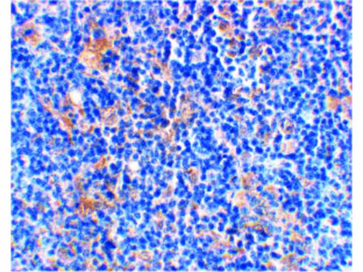
Immunohistochemistry of BAD antibody. Tissue: rat thymus tissue. Fixation: formalin fixed paraffin embedded. Antigen retrieval: not required. Primary antibody: BAD antibody at 2 ug/mL for 1 h at RT. Secondary antibody: Peroxidase rabbit secondary antibody at 1:10,000 for 45 min at RT. Localization: BAD is located in the cytoplasm, cell membrane, mitochondria, and mitochondrial outer membrane. Staining: BAD is stained with toluidine blue.
|
|
|
|
Immunofluorescence Microscopy of BAD antibody. Cell Type: rat thymus cells. Fixation: 0.5% PFA. Antigen retrieval: not required. Primary antibody: BAD antibody at 10 ug/mL for 1 h at RT. Secondary antibody: Fluorescein rabbit secondary antibody at 1:10,000 for 45 min at RT. Localization: BAD is located in the cytoplasm, cell membrane, mitochondrion, and the mitochondrial outer membrane. Staining: BAD as red fluorescent signal.
|
|
|
| メーカー |
品番 |
包装 |
|
RKL
|
200-401-Z19
|
100 UG
|
※表示価格について
| 当社在庫 |
なし
|
| 納期目安 |
約10日
|
| 保存温度 |
-20℃
|
|

