|
※サムネイル画像をクリックすると拡大画像が表示されます。
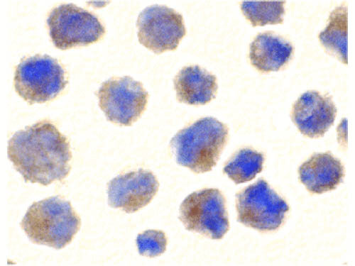
Immunocytochemistry of BACE antibody. Cells: 3T3 cells. Fixation: formaldehyde and blocked with 10% serum for 1 h at RT. Antigen retrieval: heat mediation with a citrate buffer (pH6). Primary antibody: BACE antibody at 10 μg/mL overnight at 2-8°C. Secondary antibody: Peroxidase goat anti-rabbit secondary antibody at 1:250 for 45 min at RT. Localization: BACE is localized in the cell membrane, endoplasmic reticulum, Golgi apparatus, endosome, and the cell surface. Counter stained with Hematoxylin.
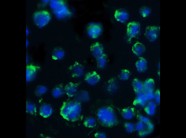
Immunofluorescence of Rabbit Anti-BACE Antibody. Cells: NIH/3T3 Cells. Fixation: 4% Paraformaldehyde. Primary Antibody: Anti-BACE at 20μg/mL. Secondary Antibody: Goat anti-Rabbit at 1:500. Staining: DAPI. Cytosol and membrane staining.
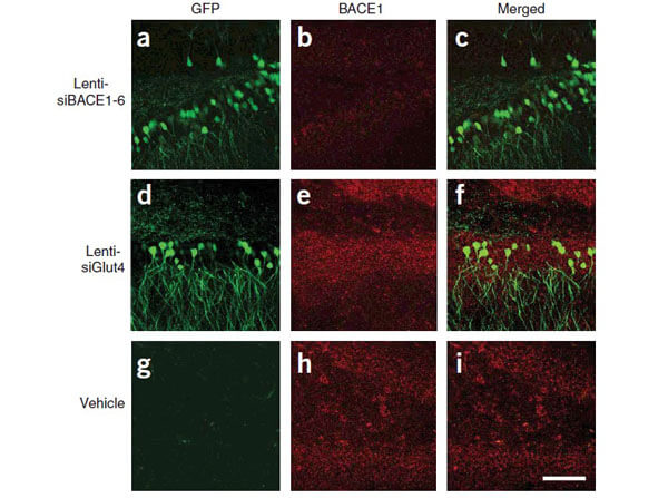
Immunofluorescence of Anti-BACE Antibody. Immunolabeling patterns of BACE1 expression and the lenti-siRNA distribution. Sections from APP transgenic mice treated with the eGFP-tagged lenti siRNA vectors (green) were co-immunolabeled with an antibody against BACE1 (red) and imaged with the LSCM. All sections are from the hippocampus of treated mice. (a?c) Lenti-siBACE1-6?treated mice. Areas within the hippocampus expressing the eGFP-tagged vector have reduced BACE1 immunolabeling. (d?f) Mice treated with the eGFP-tagged control lenti-siGlut4 show unchanged expression of BACE1 in the hippocampus. (g?i) Mice treated with a saline vehicle show unchanged expression of BACE1 in the hippocampus. (Singer et al., 2005).
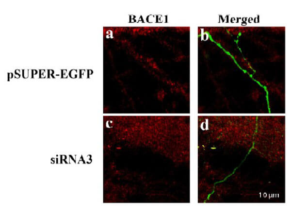
Immunofluorescence of Anti-BACE Antibody. Decreased BACE1 expression in DRG following siRNA3 transfection. DRG neurons were transfected with 1 μg siRNA3 plasmid and incubated for 48 hours in 37°C. DRG neurons were stained for BACE1 using the Anti-BACE antibody. (a,b) Neurons transfected with the control plasmid pSUPER-EGFP (green) did not display any changes in BACE1 expression (red). (c,d) DRG neurons transfected with siRNA3 displayed reduced BACE1 expression in the axon. (Hyun, 2007).
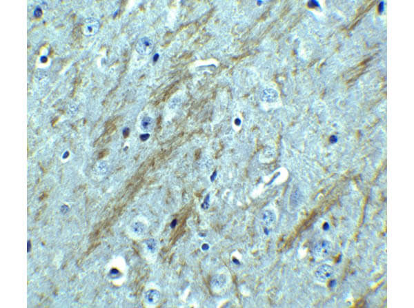
Immunohistochemistry of Rabbit anti-BACE Anitbody. Tissue: Mouse Brain. Fixation: Formaldehyde blocked with 10% serum for 1hr at RT. Antigen retrieval: heat mediation with citrate buffer pH6. Primary Antibody: Anti-BACE at 2.5μg/mL overnight at 2-8°C. Secondary Antibody: goat anti-Rabbit IgG HRP at 1:250. Counterstain: hematoxylin.
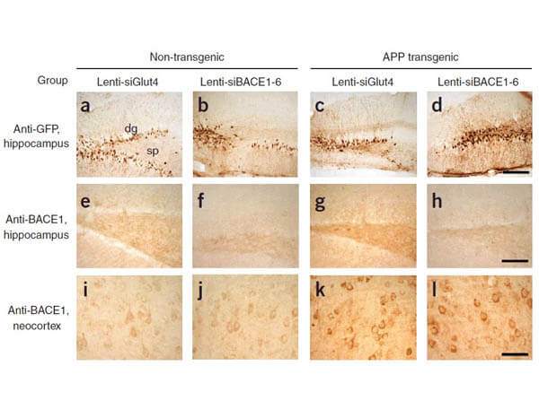
Immunohistochemistry of Rabbit Anti-BACE Antibody. Characterization of the effects of lenti-siBACE1-6 expression in the brains of APP transgenic mice. (a?d) Anti-eGFP immunoreactivity in the hippocampus (the injection site) shows comparable and consistent expression of lenti-siRNA constructs in the dentate gyrus (dg) and stratus polymorphus (sp). (e) Anti-BACE1 immunoreactivity in the hippocampus of nontransgenic mice treated with lenti-siGlut4. (f) Reduced BACE1 immunostaining in the hippocampus of nontransgenic mice treated with lenti-siBACE1-6 vector. (g) Intense BACE1 immunoreactivity in the hippocampus of APP transgenic mice treated with lenti-siGlut4. (h) Reduced BACE1 expression in APP transgenic mice treated with lenti-siBACE1-6 vector. (i,j) Anti-BACE1 reacted with pyramidal cell bodies in the neocortex, which was not injected. (Singer et al., 2005).
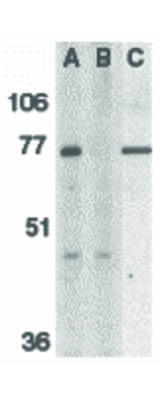
Western Blot of BACE antibody. Lane 1: Human brain tissue lysate. Lane 2: Human brain tissue lysate with the presence of blocking peptide. Lane3: Mouse 3T3 cell lysate. Load: 35 μg per lane. Primary antibody: BACE antibody at 1 μg/mL for overnight at 4°C. Secondary antibody: Peroxidase rabbit secondary antibody at 1:10,000 for 45 min at RT. Block: 5% BLOTTO overnight at 4°C. Predicted/Observed size: 55.7 kDa, 76 kDa for BACE. Other band(s): BACE splice variants and isoforms.
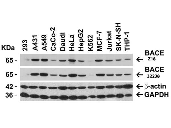
Western Blot of Anti-BACE Antibody. Loading: 15 μg of lysates per lane. Primary Antibodies: Anti-BACE 200-401-Z18 (1μg/mL), BACE (1μg/mL), beta-actin (1μg/mL), and GAPDH (0.02μg/mL), for 1hr at RT in 5% NFDM/TBST. Secondary Antibody: Goat anti-rabbit IgG HRP conjugate at 1:10,000.
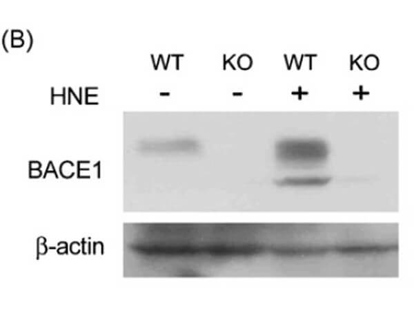
Western Blot of Anti-BACE Antibody. Wildtype and BACE −/− MEFs were exposed to HNE (15_M) for 2 hr. BACE1 levels were examined by Western blot with anti-BACE antibodies. (Jo et al., 2010)
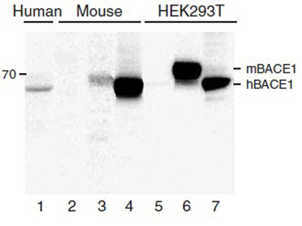
Western Blot of Anti-BACE Antibody. Lanes 1?4 are frontal cortex homogenates from human and mouse brains. Lane 1 is from a neurologically unimpaired aged human control case, lane 2 from a BACE1-deficient mouse, lane 3 from a nontransgenic mouse and lane 4 from hBACE1 transgenic mouse. Lanes 5?7 are lysates from HEK293T cells transfected with a plasmid vector expressing eGFP, mBACE1 and hBACE1, respectively. BACE1 antibody recognized both the mouse and human forms of BACE1. (Singer et al., 2005).
|
|
|
|
Immunocytochemistry of BACE antibody. Cells: 3T3 cells. Fixation: formaldehyde and blocked with 10% serum for 1 h at RT. Antigen retrieval: heat mediation with a citrate buffer (pH6). Primary antibody: BACE antibody at 10 μg/mL overnight at 2-8°C. Secondary antibody: Peroxidase goat anti-rabbit secondary antibody at 1:250 for 45 min at RT. Localization: BACE is localized in the cell membrane, endoplasmic reticulum, Golgi apparatus, endosome, and the cell surface. Counter stained with Hematoxylin.
|
|
| 別品名 |
BACE Antibody, ASP2, BACE, HSPC104, KIAA1149, Beta-secretase 1, Aspartyl protease 2, ASP2
|
| 交差種 |
Human
Mouse
|
| 適用 |
Western Blot
Enzyme Linked Immunosorbent Assay
Immunohistochemistry
Immuno Fluorescence
|
| 免疫動物 |
Rabbit
|
| 抗原部位 |
C-terminus
|
| 標識物 |
Unlabeled
|
| 精製度 |
Ig fraction - Ion Exchange /Gel Filtration
|
| GENE ID |
23621
|
| Accession No.(Gene/Protein) |
AF190725, P56817
|
| Gene Symbol |
BACE1
|
| 参考文献 |
[Pub Med ID]20854419
|
| [注意事項] |
濃度はロットによって異なる可能性があります。メーカーDS及びCoAからご確認ください。
|
|
| メーカー |
品番 |
包装 |
|
RKL
|
200-401-Z18
|
100 UG
|
※表示価格について
| 当社在庫 |
なし
|
| 納期目安 |
約10日
|
| 保存温度 |
-20℃
|
|
※当社では商品情報の適切な管理に努めておりますが、表示される法規制情報は最新でない可能性があります。
また法規制情報の表示が無いものは、必ずしも法規制に非該当であることを示すものではありません。
商品のお届け前に最新の製品法規制情報をお求めの際はこちらへお問い合わせください。
|
※当社取り扱いの試薬・機器製品および受託サービス・創薬支援サービス(納品物、解析データ等)は、研究用としてのみ販売しております。
人や動物の医療用・臨床診断用・食品用としては、使用しないように、十分ご注意ください。
法規制欄に体外診断用医薬品と記載のものは除きます。
|
|
※リンク先での文献等のダウンロードに際しましては、掲載元の規約遵守をお願いします。
|
|
※CAS Registry Numbers have not been verified by CAS and may be inaccurate.
|
