|
※サムネイル画像をクリックすると拡大画像が表示されます。
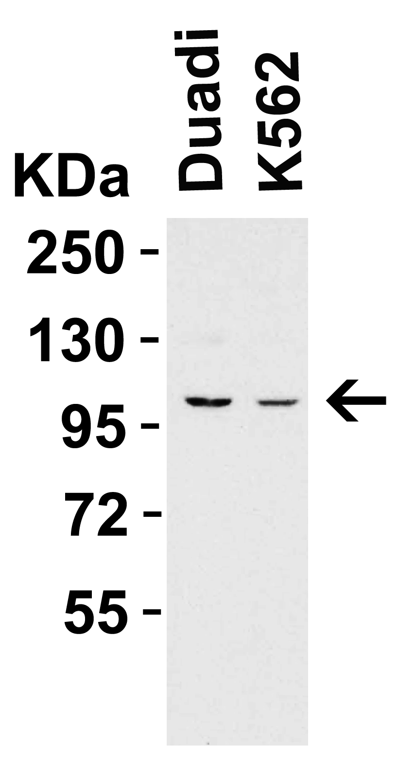
Figure 1 WB Validation in Human Cell Lines
Loading: 15 μg of human cell lysate Antibodies: PPARGC1A 7793, 2 μg/mL , 1 h incubation at RT in 5% NFDM/TBST. Secondary: Goat Anti-Rabbit IgG HRP conjugate at 1:10000 dilution.
WB Validation in Human Lung
Loading: 15 μg of human lung lysate Antibodies: PPARGC1A 7793, 2 μg/mL , 1 h incubation at RT in 5% NFDM/TBST. Secondary: Goat Anti-Rabbit IgG HRP conjugate at 1:10000 dilution.
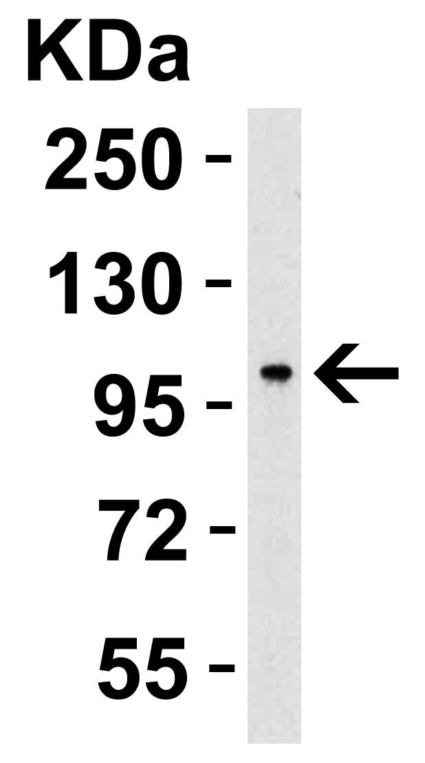
Figure 3 WB Validation in Mouse Skin
Loading: 15 μg of mouse skin lysate Antibodies: PPARGC1A 7793, 2 μg/mL , 1 h incubation at RT in 5% NFDM/TBST. Secondary: Goat Anti-Rabbit IgG HRP conjugate at 1:10000 dilution.
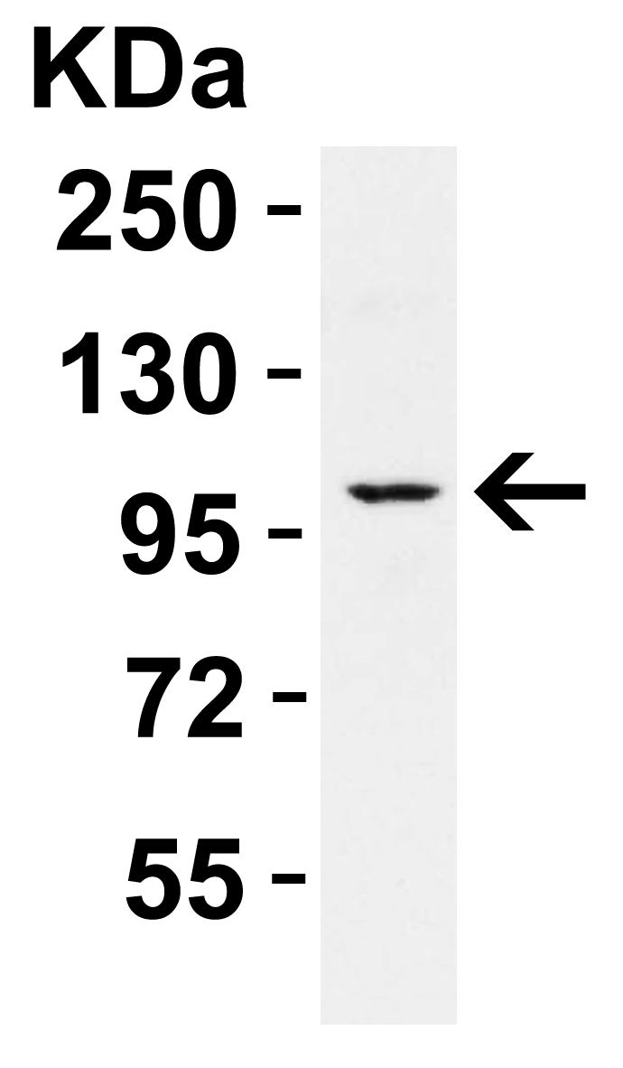
Figure 4 WB Validation in Rat Kidney
Loading: 15 μg of rat kidney lysate Antibodies: PPARGC1A 7793, 2 μg/mL , 1 h incubation at RT in 5% NFDM/TBST. Secondary: Goat Anti-Rabbit IgG HRP conjugate at 1:10000 dilution.

Figure 5 Immunofluorescence Validation of PPARGC1A in MCF7 Cells
Immunofluorescent analysis of 4% paraformaldehyde-fixed MCF7 cells labeling PPARGC1A with 7793 at 20 μg/mL, followed by goat anti-rabbit IgG secondary antibody at 1/500 dilution (green) and DAPI staining (blue).
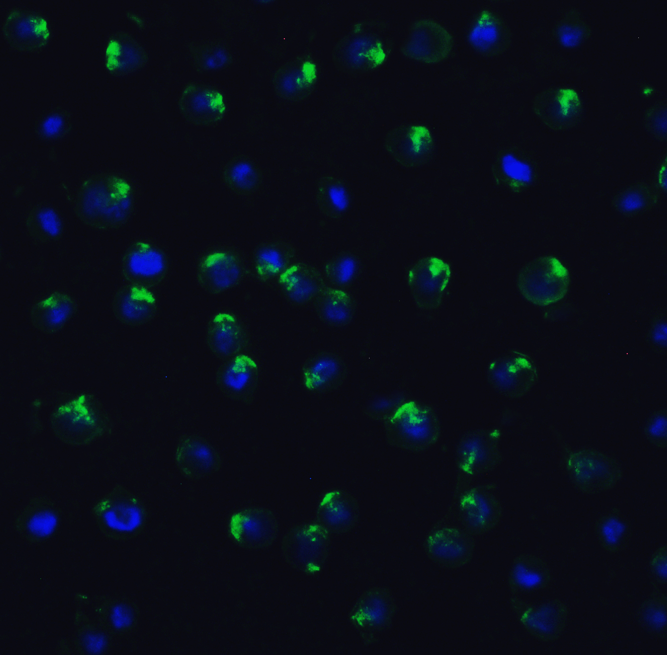
Figure 6 Immunofluorescence Validation of PPARGC1A in HELA Cells
Immunofluorescent analysis of 4% paraformaldehyde-fixed HeLa cells labeling PPARGC1A with 7793 at 20 μg/mL, followed by goat anti-rabbit IgG secondary antibody at 1/500 dilution (green) and DAPI staining (blue).
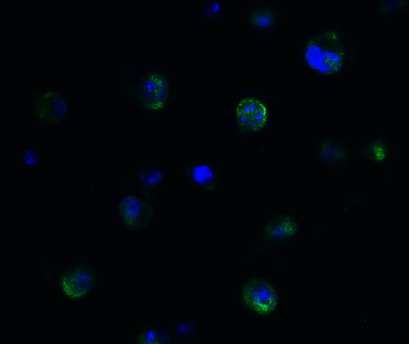
Figure 7 Immunohistochemistry Validation of PPARGC1A in Human Colon Carcinoma Tissue
Immunohistochemical analysis of paraffin-embedded human colon carcinoma tissue using anti-PPARGC1A antibody (7793) at 1 μg/ml. Tissue was fixed with formaldehyde and blocked with 10% serum for 1 h at RT; antigen retrieval was by heat mediation with a citrate buffer (pH6). Samples were incubated with primary antibody overnight at 4℃. A goat anti-rabbit IgG H&L (HRP) at 1/250 was used as secondary. Counter stained with Hematoxylin.
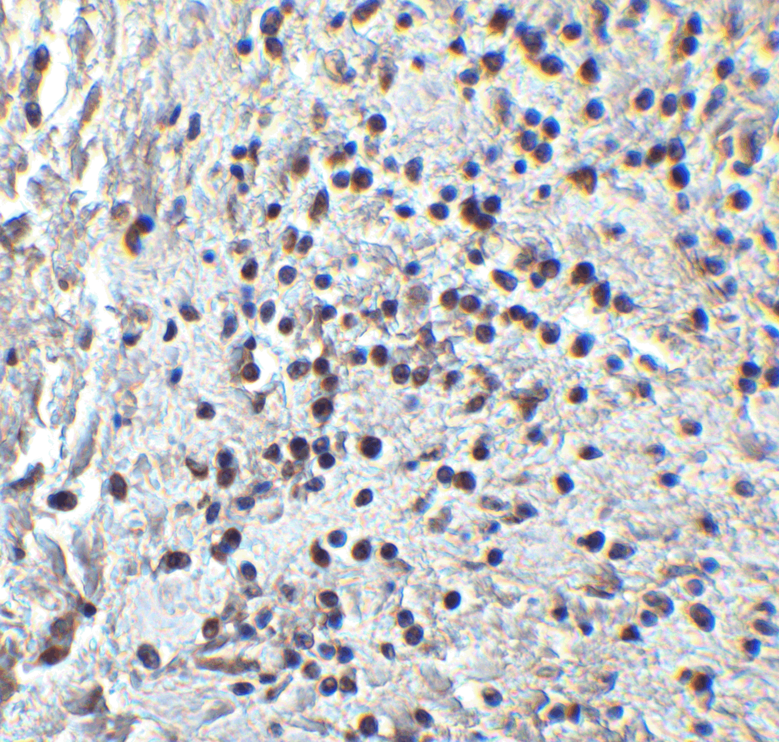
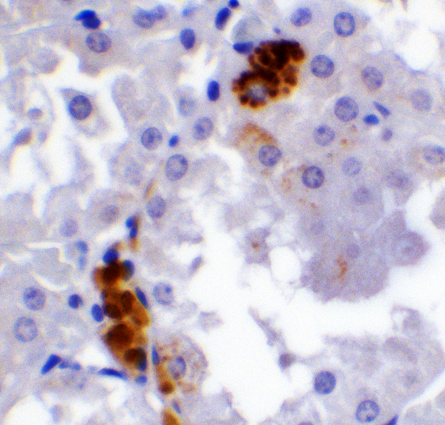
Figure 8 Immunohistochemistry Validation of PPARGC1A in Human Pancreas Tissue
Immunohistochemical analysis of paraffin-embedded human pancreas tissue using anti-PPARGC1A antibody (7793) at 2 μg/ml. Tissue was fixed with formaldehyde and blocked with 10% serum for 1 h at RT; antigen retrieval was by heat mediation with a citrate buffer (pH6). Samples were incubated with primary antibody overnight at 4℃. A goat anti-rabbit IgG H&L (HRP) at 1/250 was used as secondary. Counter stained with Hematoxylin.
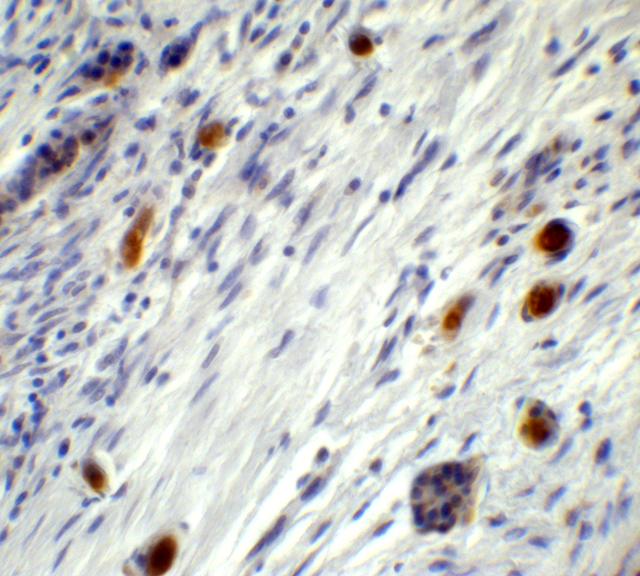
Figure 9 Immunohistochemistry Validation of PPARGC1A in Mouse Stomach Tissue
Immunohistochemical analysis of paraffin-embedded mouse stomach tissue using anti-PPARGC1A antibody (7793) at 2 μg/ml. Tissue was fixed with formaldehyde and blocked with 10% serum for 1 h at RT; antigen retrieval was by heat mediation with a citrate buffer (pH6). Samples were incubated with primary antibody overnight at 4℃. A goat anti-rabbit IgG H&L (HRP) at 1/250 was used as secondary. Counter stained with Hematoxylin.

Figure 10 Immunohistochemistry Validation of PPARGC1A in Rat Kidney Tissue
Immunohistochemical analysis of paraffin-embedded rat kidney tissue using anti-PPARGC1A antibody (7793) at 2 μg/ml. Tissue was fixed with formaldehyde and blocked with 10% serum for 1 h at RT; antigen retrieval was by heat mediation with a citrate buffer (pH6). Samples were incubated with primary antibody overnight at 4℃. A goat anti-rabbit IgG H&L (HRP) at 1/250 was used as secondary. Counter stained with Hematoxylin.
|
|
|
|
Figure 1 WB Validation in Human Cell Lines
Loading: 15 μg of human cell lysate Antibodies: PPARGC1A 7793, 2 μg/mL , 1 h incubation at RT in 5% NFDM/TBST. Secondary: Goat Anti-Rabbit IgG HRP conjugate at 1:10000 dilution.
|
|
| 別品名 |
PPARGC1A Antibody: LEM6, PGC1, PGC1A, PGC-1v, PPARGC1, PGC-1(alpha), LEM6, Peroxisome proliferator-activated receptor gamma coactivator 1-alpha, Ligand effect modulator 6, PGC-1-alpha
|
| 交差種 |
Human
Mouse
Rat
|
| 適用 |
Western Blot
IHC paraffin embedding section
Enzyme Linked Immunosorbent Assay
Immuno Fluorescence
|
| 免疫動物 |
Rabbit
|
| 抗体クラス |
IgG
|
| 抗原部位 |
N-terminus
|
| 標識物 |
Unlabeled
|
| 精製度 |
Affinity Purified
|
| GENE ID |
10891
|
| Accession No.(Gene/Protein) |
NP_037393
|
| Gene Symbol |
PPARGC1A
|
| 推奨品 |
ポジティブコントロール 品番:1301 - Human Heart Tissue Lysate
|
| その他 |
[Protein GI Number]7019499
[Swiss-Prot No]Q9UBK2
|
| 参考文献 |
Tsuemi T and La Spada AR. PGC-1a at the intersection of bioenergetics regulation and neuron function: from Huntington's disease to Parkinson's disease and beyond. Prog. Neurobiol. 2012; 97:142-51.
Kang C and Li Ji L. Role of PGC-1a signaling in skeletal muscle health and disease. Ann. NY Acad. Sci. 2012; 1271:110-7.
Liu C and Lin JD. PGC-1 coactivators in the control of energy metabolism. Acta Biochim. Biophys. Sin. 2011; 43:248-57.
|
|
| メーカー |
品番 |
包装 |
|
PSC
|
7793
|
20 UG
[1 mg/mL]
|
※表示価格について
| 当社在庫 |
なし
|
| 納期目安 |
3週間程度
|
| 保存温度 |
-20℃
|
|
※当社では商品情報の適切な管理に努めておりますが、表示される法規制情報は最新でない可能性があります。
また法規制情報の表示が無いものは、必ずしも法規制に非該当であることを示すものではありません。
商品のお届け前に最新の製品法規制情報をお求めの際はこちらへお問い合わせください。
|
※当社取り扱いの試薬・機器製品および受託サービス・創薬支援サービス(納品物、解析データ等)は、研究用としてのみ販売しております。
人や動物の医療用・臨床診断用・食品用としては、使用しないように、十分ご注意ください。
法規制欄に体外診断用医薬品と記載のものは除きます。
|
|
※リンク先での文献等のダウンロードに際しましては、掲載元の規約遵守をお願いします。
|
|
※CAS Registry Numbers have not been verified by CAS and may be inaccurate.
|
