
| 別品名 |
CXCR4 Antibody: FB22, HM89, LAP3, LCR1, NPYR, WHIM, CD184, LESTR, NPY3R, NPYRL, HSY3RR, NPYY3R, D2S201E
|
| 種由来 |
Human
|
| 標識物 |
Unlabeled
|
| 適用 |
Western Blot
Enzyme Linked Immunosorbent Assay
Immunohistochemistry
Immuno Fluorescence
|
| 免疫動物 |
Rabbit
|
| 抗体クラス |
IgG
|
| 交差種 |
Human
Mouse
|
| GENE ID |
7852
|
| Accession No.(Gene/Protein) |
NP_003458
|
| Gene Symbol |
CXCR4
|
| 形状 |
液状
|
| 推奨品 |
ポジティブコントロール 品番:1201 - HeLa Cell Lysate
|
| その他 |
[Protein GI Number]4503175
[Swiss-Prot No]P61073
|
| 参考文献 |
Dimitrov DS. Cell 1997;91:721-730
Feng Y et al. Science 1996;272:872-7
Berson JF et al. J Virol 1996;70:6288-95
Doranz BJ et al. Cell 1996;85:1149-1158
|
|
※サムネイル画像をクリックすると拡大画像が表示されます。
Figure 1 Western Blot Validation of CXCR4 in HeLa Cells
Loading: 15 μg of lysates per lane.Antibodies: 1012 (1 μg/mL), 1 h incubation at RT in 5% NFDM/TBST. Secondary: Goat anti-rabbit IgG HRP conjugate at 1:10000 dilution.
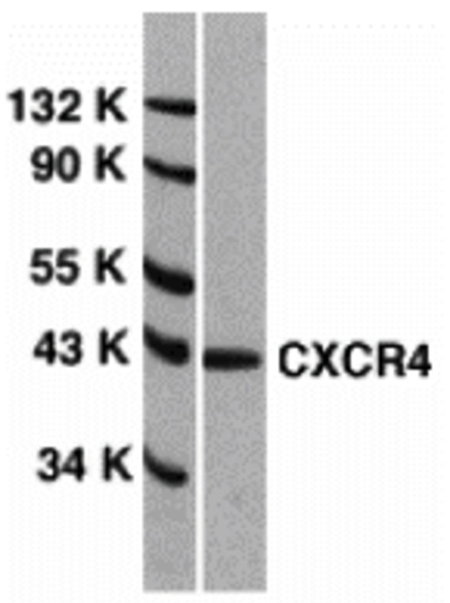
Figure 2 Independent Antibody Validation (IAV) via Protein Expression Profile
Loading: 15 μg of lysates per lane. Antibodies: 1009 (1 μg/mL), 1012 (1 μg/mL), and beta-actin (1 μg/mL), 1 h incubation at RT in 5% NFDM/TBST. Secondary: Goat anti-rabbit IgG HRP conjugate at 1:10000 dilution.
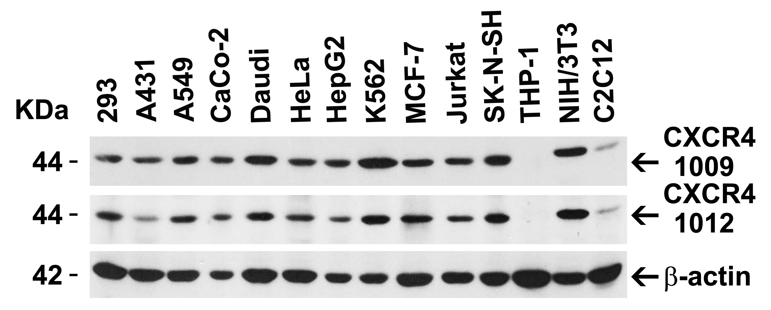
Figure 3 Validation with CXCR4 siRNA Knockdown in HeLa Cells
HeLa cells were transfected with control siRNAs (lane 1) or CXCR4 siRNAs (lane 2) Loading: 10 μg of HeLa whole cell lysates per lane. Antibodies: 1012 (2 μg/mL), 1 h incubation at RT in 5% NFDM/TBST. Secondary: Goat anti-rabbit IgG HRP conjugate at 1:10000 dilution.
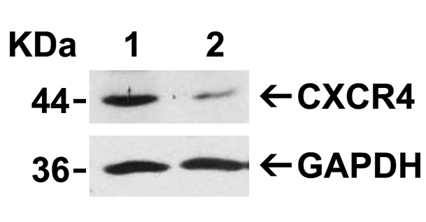
Figure 4 Animal Species Reactivity
Loading: Lysates/proteins at 20 μg per lane.Antibodies: 1009 (2 μg/mL) or 1012 (2 μg/mL). 1 h incubation at RT in 5% NFDM/TBST.Secondary: Goat anti-rabbit IgG HRP conjugate at 1:10000 dilution.
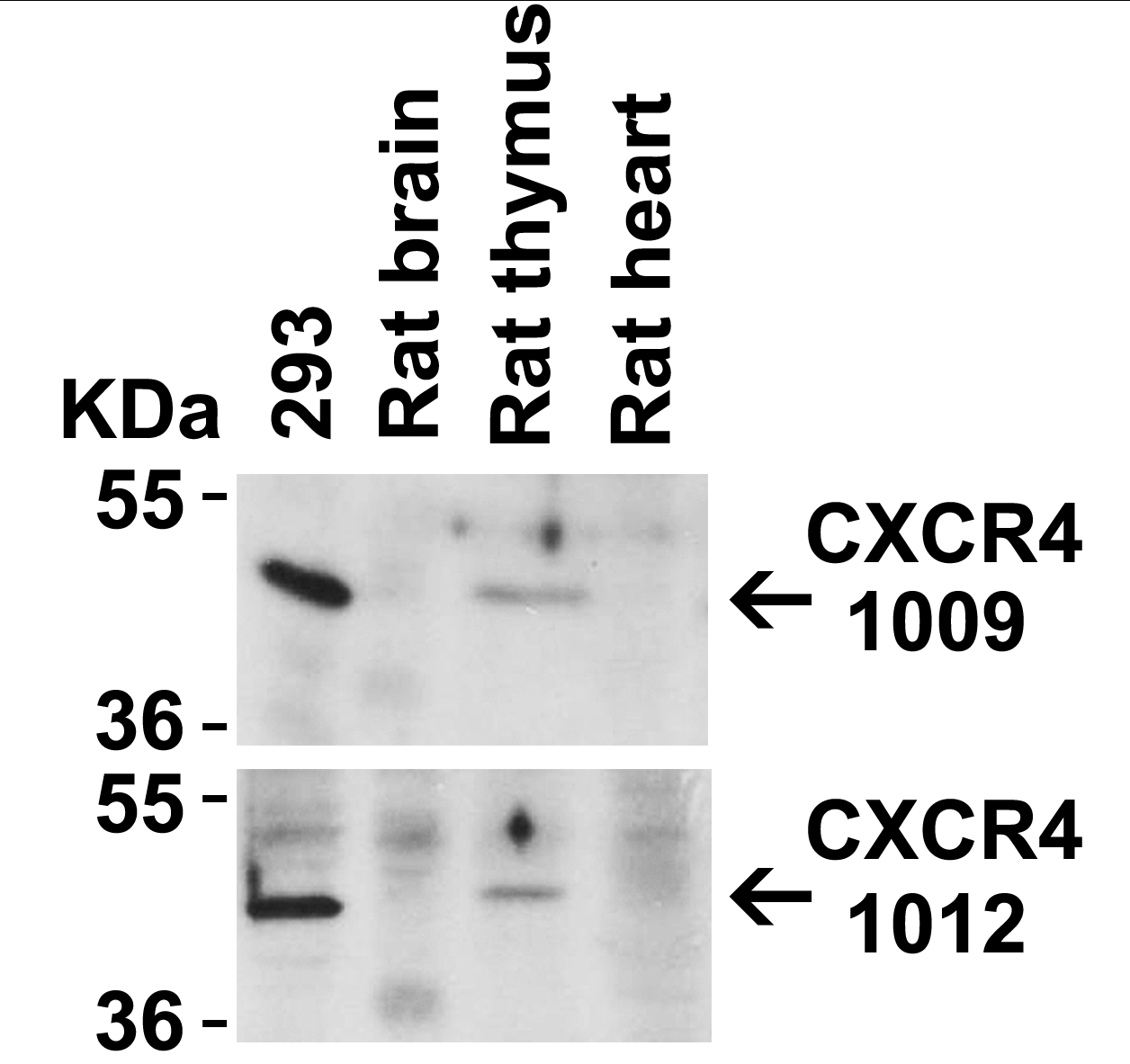
Figure 5 Immunofluorescence Validation of CXCR4
Immunofluorescent analysis of 4% paraformaldehyde-fixed HeLa cells labeling CXCR4 with 1012 at 4 μg/mL, followed by goat anti-rabbit IgG secondary antibody at 1/500 dilution (red). Image showing both membrane and cytoplasmic staining on HeLa cell line.
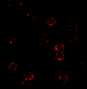
Figure 6 Immunohistochemistry Validation of CXCR4 in Human Spleen
Immunohistochemical analysis of paraffin-embedded human spleen tissue using anti-CXCR4 antibody (1012) at 5 μg/ml. Tissue was fixed with formaldehyde and blocked with 10% serum for 1 h at RT; antigen retrieval was by heat mediation with a citrate buffer (pH6). Samples were incubated with primary antibody overnight at 4℃. A goat anti-rabbit IgG H&L (HRP) at 1/250 was used as secondary. Counter stained with Hematoxylin.
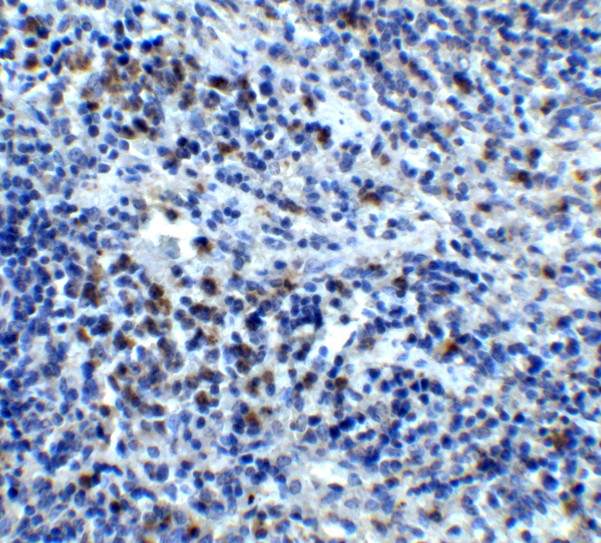
Figure 7 KO Validation of CXCR4 by Flow Cytometry (Odemis, et al., 2010)
Astrocytes from wild-type or CXCR4 knockout mice were stained with primary antibodies against CXCR4 and FITC-labeled secondary antibodies, and subsequently subjected to flow cytometry. CXCR4−/− astrocytes (red) showed loss of CXCR4 cell-surface expression compared with wild-type cells (black).
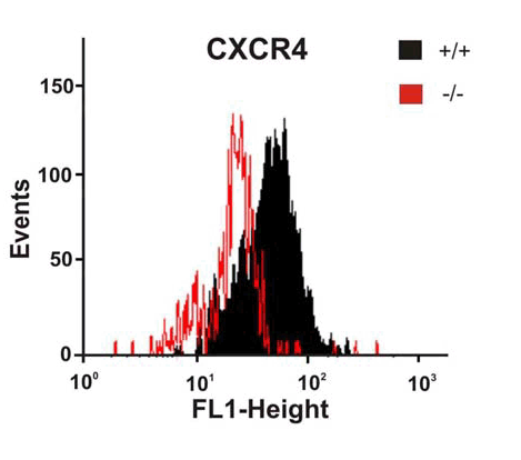
Figure 8 Overexpression Validation of CXCR4 (Kozak et al., 2002)
U87MG and U87MG-CXCR4 extracts were included as negative and positive controls, respectively, for CXCR4 detection with anti-CXCR4 antibodies.
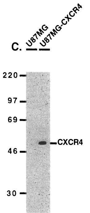
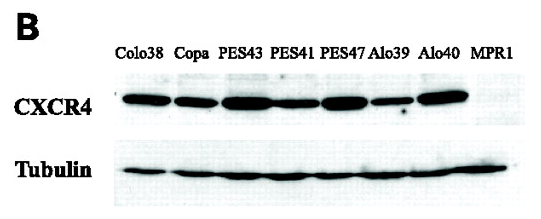
Figure 9 WB Validation of CXCR4 in Human Metastatic Melanoma (Scala et al., 2006)
CXCR4 protein was detected in the human metastatic melanoma cell lines and human melanoma cell line (colo38), but not in the human primary melanocytes (MPR1) with anti-CXCR4 antibodies.
|
|
|
|
Figure 1 Western Blot Validation of CXCR4 in HeLa Cells
Loading: 15 μg of lysates per lane.Antibodies: 1012 (1 μg/mL), 1 h incubation at RT in 5% NFDM/TBST. Secondary: Goat anti-rabbit IgG HRP conjugate at 1:10000 dilution.
|
|
|
| メーカー |
品番 |
包装 |
|
PSC
|
1012
|
20 UG
[1 mg/mL]
|
※表示価格について
| 当社在庫 |
なし
|
| 納期目安 |
3週間程度
|
| 保存温度 |
-20℃
|
|

