|
※サムネイル画像をクリックすると拡大画像が表示されます。
|
|
|
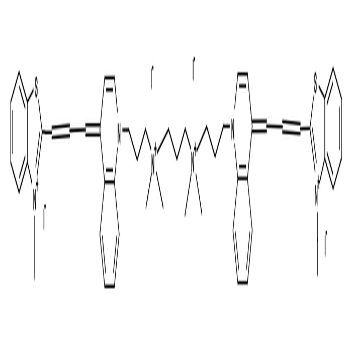
| 励起波長 |
642
|
| 蛍光波長 |
660
|
| 分子量 |
1354.85
|
| 参考文献 |
Bonaccorsi S, Giansanti MG, Cenci G, Gatti M.. (2012). Chromatin staining of Drosophila testes. Cold Spring Harb Protoc, doi:10.1101/pdb.prot067363.
Cho H, Indig GL, Weichert J, Shin HC, Kwon GS.. (2012). In vivo cancer imaging by poly(ethylene glycol)-b-poly(varepsilon-caprolactone) micelles containing a near-infrared probe. Nanomedicine, 8, 228.
Henneberger R, Birch D, Bergquist P, Walter M, Anitori RP.. (2011). The fluorescent dyes TO-PRO-3 and TOTO-3 iodide allow detection of microbial cells in soil samples without interference from background fluorescence. Biotechniques, 51, 190.
Haberl S, Miklavcic D, Pavlin M.. (2010). Effect of Mg ions on efficiency of gene electrotransfer and on cell electropermeabilization. Bioelectrochemistry, 79, 265.
Glaser K, Wilke K, Wepf R, Biel SS.. (2008). Visualizing nuclei in skin cryosections: viable options to 4'6-diamidino-2-phenylindol for confocal laser microscopy. Skin Res Technol, 14, 324.
Phe MH, Dossot M, Guilloteau H, Block JC.. (2007). Highly chlorinated Escherichia coli cannot be stained by propidium iodide. Can J Microbiol, 53, 664.
Martin RM, Leonhardt H, Cardoso MC.. (2005). DNA labeling in living cells. Cytometry A, 67, 45.
Shan L.. (2004). Quinolinium, 1,1'-[1,3-propanediylbis[(dimethyliminio)-3,1-propanediyl]]bis[4-[3-(3-methyl-2(3 H)-benzothiazolylidene)-1-propen-1-yl]-,iodide (1:4). In: Molecular Imaging and Contrast Agent Database (MICAD), Bethesda (MD)., .
Phe MH, Dossot M, Block JC.. (2004). Chlorination effect on the fluorescence of nucleic acid staining dyes. Water Res, 38, 3729.
Luppens SB, Barbaras B, Breeuwer P, Rombouts FM, Abee T.. (2003). Selection of fluorescent probes for flow cytometric viability assessment of Listeria monocytogenes exposed to membrane-active and oxidizing disinfectants. J Food Prot, 66, 1393.
Zuliani T, Duval R, Jayat C, Schnebert S, Andre P, Dumas M, Ratinaud MH.. (2003). Sensitive and reliable JC-1 and TOTO-3 double staining to assess mitochondrial transmembrane potential and plasma membrane integrity: interest for cell death investigations. Cytometry A, 54, 100.
Golzio M, Teissie J, Rols MP.. (2002). Direct visualization at the single-cell level of electrically mediated gene delivery. Proc Natl Acad Sci U S A, 99, 1292.
Bunthof CJ, Bloemen K, Breeuwer P, Rombouts FM, Abee T.. (2001). Flow cytometric assessment of viability of lactic acid bacteria. Appl Environ Microbiol, 67, 2326.
Boutonnat J, Barbier M, Ronot X, Seigneurin D.. (2000). Nucleus labeling or membrane labeling for studying the proliferation of drug treated cells. Morphologie, 84, 11.
Kahn E, Lizard G, Frouin F, Roignot P, Chardonnet Y, Di Paola R.. (2000). Factor analysis of confocal image sequences of human papillomavirus DNA revealed with fast red in cervical tissue sections stained with TOTO-iodide. Anal Quant Cytol Histol, 22, 168.
|
| 使用文献 |
Bonaccorsi S Giansanti MG, Cenci G, Gatti M. (2012). Chromatin staining of Drosophila testes. Cold Spring Harb Protoc.
|
|
| メーカー |
品番 |
包装 |
|
ABD
|
17576
|
0.2 ML
|
※表示価格について
| 当社在庫 |
なし
|
| 納期目安 |
1週間程度
|
| 法規制 |
安・危
|
| 保存温度 |
-20℃
|
|